
Loading Diagnostics...
Connecting to our secure server
Connecting to our secure server

Browse our comprehensive library of diagnostic capabilities. We provide over 200 NABL accredited radiology scans and automated clinical pathology investigations.
Because accurate diagnosis leads to better treatment, advanced Radiology Services empower clinical evaluations through high-resolution MRI, multi-slice CT, ultrasound, Doppler, digital mammography, X-ray, PET-CT, DEXA, and nuclear medicine.
Fundamentally, professional Radiology Services represent the core foundation of modern clinical diagnostics, using non-invasive imaging technologies to capture internal anatomical, structural, and functional details of the human body. Consequently, by utilizing magnetic fields, sound waves, ionizing radiation, and radiopharmaceuticals, diagnostic imaging enables physicians to accurately identify diseases, assess trauma, monitor organ function, and evaluate treatment efficacy.
Moreover, comprehensive Radiology Services play a vital role across all medical specialties, including neurology, cardiology, oncology, orthopedics, gastroenterology, and obstetrics. Therefore, from detecting microscopic tissue anomalies to guiding precision interventional procedures, specialized imaging ensures evidence-based medical management and enhanced patient safety.
Specifically, medical imaging encompasses distinct diagnostic modalities tailored to evaluate specific anatomical structures and physiological states. As a result, below is an overview of the primary imaging modalities utilized in modern Radiology Services:
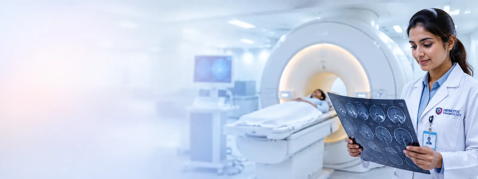
Additionally, Magnetic Resonance Imaging provides multiplanar, high-resolution soft tissue visualization without ionizing radiation, ideal for brain, spine, joint, and pelvic evaluations.
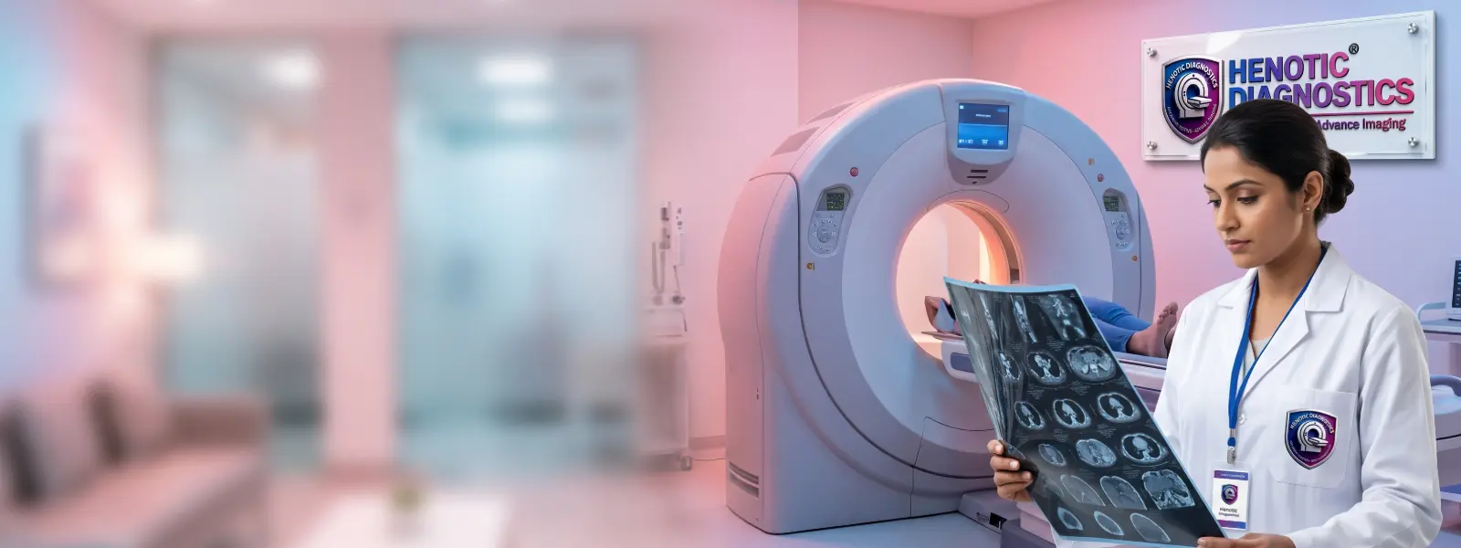
Furthermore, Computed Tomography delivers cross-sectional structural imaging of bone, chest, abdominal organs, and vascular networks with rapid multi-slice acquisition.

In addition, radiation-free imaging utilizes sound waves for real-time visualization of abdominal organs, pelvic structures, soft tissues, and obstetric growth monitoring.
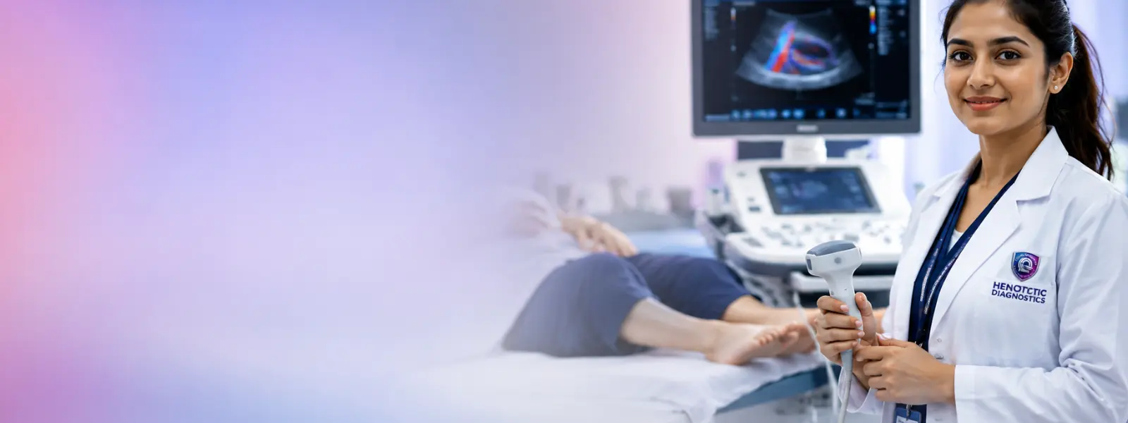
Similarly, specialized hemodynamic imaging evaluates arterial and venous blood flow velocity, vascular resistance, and hemodynamic perfusion in real time.

Likewise, fundamental digital radiography provides immediate assessment of skeletal integrity, pulmonary conditions, joint alignment, and abdominal emergencies.
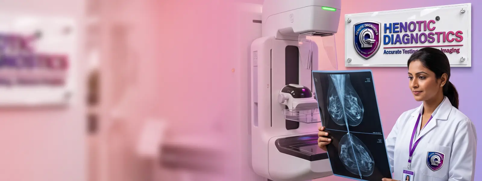
Besides, low-dose digital mammography and sonomammography are engineered for early detection of microcalcifications, masses, and breast tissue irregularities.

Equally important, integrated Positron Emission Tomography and CT provide metabolic, cellular-level functional insights merged with anatomical localization.
Correspondingly, physiological imaging uses radiotracers to evaluate organ function, bone turnover, thyroid metabolism, and neuroendocrine activity.
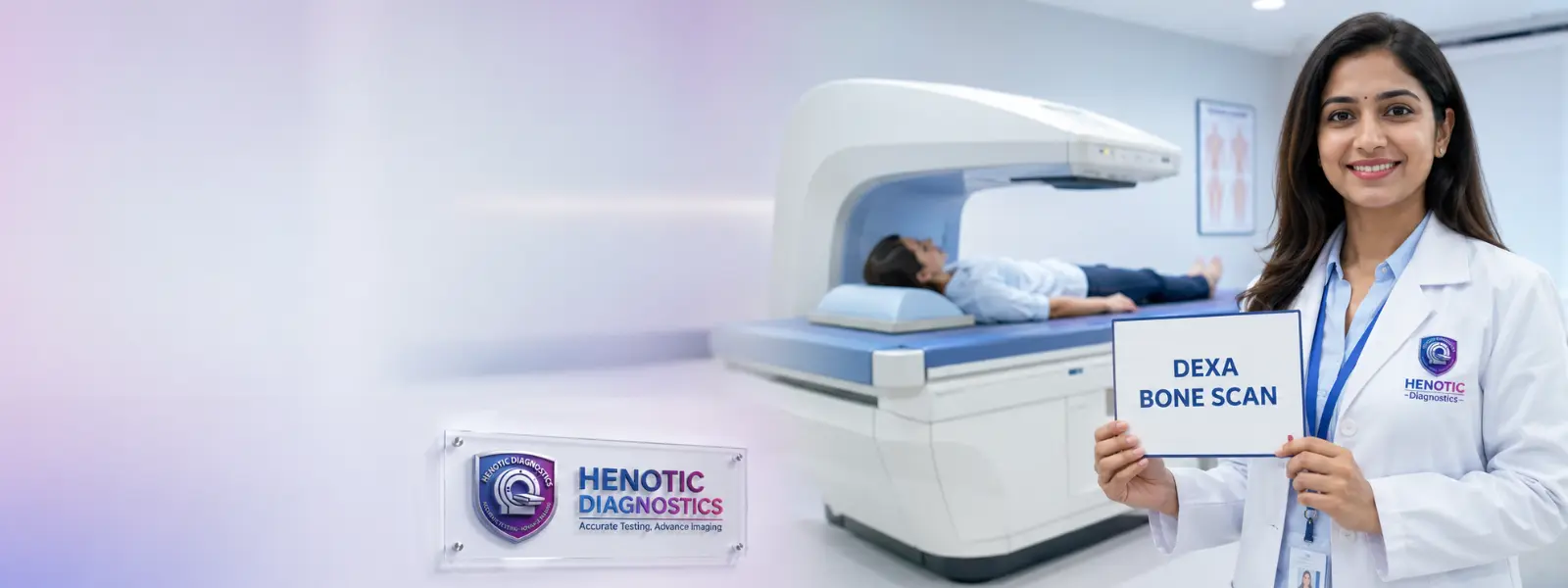
Finally, Dual-Energy X-Ray Absorptiometry offers precise quantification of bone mineral density (BMD) for diagnosing osteoporosis and predicting fracture risks.
Undeniably, Magnetic Resonance Imaging (MRI) employs powerful magnetic fields and radiofrequency pulses to generate cross-sectional images with high soft-tissue contrast resolution. Therefore, MRI represents a cornerstone of non-invasive Radiology Services for neuroimaging, musculoskeletal assessment, and pelvic pathology identification.
Specifically, detailed evaluation of cerebral parenchyma, stroke identification, demyelinating conditions, cranial nerve pathology, and intracranial lesions.
Furthermore, high-definition visualization of cervical, thoracic, and lumbar intervertebral discs, spinal cord signal intensity, and nerve root compression.
In addition, comprehensive soft-tissue diagnosis for meniscus tears, ACL/PCL ligament injuries, tendonitis, articular cartilage loss, and joint effusions.
Moreover, detailed multiplanar imaging of rotator cuff tears, labral lesions, shoulder impingement syndrome, and soft tissue masses.
Additionally, cross-sectional multiparametric characterization of focal liver lesions, pancreatic ductal disease, renal parenchyma, fibroids, and endometriosis.
As a result, non-invasive fluid-sensitive evaluation of intrahepatic biliary ducts, common bile duct stones (choledocholithiasis), and pancreatic ducts.
Consequently, radiation-free contrast and non-contrast mapping of intracranial cerebral arteries, carotid bifurcations, and renal arteries.
Finally, advanced protocols including Diffusion-Weighted Imaging (DWI), MR Spectroscopy (MRS), functional MRI (fMRI), and cardiac viability assessment.
Similarly, Computed Tomography (CT) combines specialized rotating X-ray tubes with digital detector arrays to construct detailed axial, sagittal, and coronal slice images. Consequently, advanced multi-detector CT protocols facilitate rapid volumetric Radiology Services for complex anatomical regions.
For example, rapid cross-sectional evaluation for acute intracranial hemorrhage, ischemic infarction, traumatic skull fractures, and cerebral edema.
For instance, High-Resolution Computed Tomography (HRCT) for evaluating interstitial lung disease (ILD), pulmonary fibrosis, and bronchiectasis.
In particular, multi-phase contrast imaging for acute appendicitis, diverticulitis, bowel obstruction, liver metastases, and abdominal trauma.
Specifically, unenhanced low-dose volumetric protocol providing high sensitivity for renal calculi, ureteric stones, and obstructive uropathy.
Additionally, 3D arterial vascular mapping for thoracic/abdominal aortic aneurysms, pulmonary embolism (CTPA), and peripheral vascular stenosis.
Thus, ECG-gated non-invasive visualization of coronary artery lumen, atherosclerotic plaque composition, and Calcium Scoring.
Indeed, diagnostic ultrasound utilizes high-frequency acoustic waves to generate real-time dynamic images without ionizing radiation. Because it is completely safe, acoustic scanning represents versatile Radiology Services for abdominal, pelvic, small-part, and obstetric conditions.
Firstly, evaluation of liver echogenicity, gallstones (cholelithiasis), splenomegaly, pancreatic structure, and renal parenchymal thickness.
Secondly, transabdominal and transvaginal sonography for uterine leiomyomas, ovarian cysts, endometrial thickness, and pelvic inflammatory disease.
Thirdly, targeted renal assessment for hydronephrosis, renal cysts, bladder wall thickening, and post-void residual urine volume.
Next, high-resolution sonography for thyroid nodule stratification (TI-RADS), parathyroid adenomas, and cervical lymph node evaluation.
Furthermore, targeted acoustic assessment to differentiate solid breast masses from fluid-filled benign cysts in dense tissue.
In addition, comprehensive fetal gestational tracking, nuchal translucency (NT) screening, anomaly scans, and fetal biometry.
Finally, dynamic acoustic imaging for superficial lipomas, tendon tears, joint effusions, and subcutaneous abscesses.
Because vascular health requires precise hemodynamic evaluation, Color Doppler ultrasound measures frequency shifts of reflected acoustic waves from moving blood cells. Therefore, these specialized Radiology Services provide critical diagnostic data regarding vessel patency, flow velocity, turbulent blood flow, and stenosis severity.
Specifically, assessment of carotid artery intima-media thickness (CIMT) and atheromatous plaque burden to stratify stroke risk.
Additionally, arterial and venous flow evaluation for Deep Vein Thrombosis (DVT), chronic venous insufficiency, and peripheral arterial occlusion.
Moreover, vascular flow assessment for subclavian steal syndrome, thoracic outlet arterial compression, and upper extremity thrombosis.
Furthermore, hemodynamic Doppler waveform analysis and Ankle-Brachial Index (ABI) correlation for peripheral artery disease (PAD).
Consequently, transcranial Doppler imaging measuring fetal MCA peak systolic velocity to detect fetal anemia and intracranial hemodynamics.
As a result, renal artery flow velocity measurement and resistive index (RI) calculation to diagnose renovascular hypertension.
In addition, placental resistance Doppler monitoring for intrauterine growth restriction (IUGR) and pre-eclampsia risk assessment.
Finally, comprehensive systemic color Doppler mapping for vascular malformations, varicose veins, and aneurysm flow patterns.
Additionally, Undoubtedly, digital radiography (X-ray) remains the foundational diagnostic imaging modality, offering rapid, low-dose examination of skeletal structures, joint articulations, and thoracic cavity organs within modern Radiology Services.
For example, standard PA and lateral projections for pneumonia, cardiomegaly, pneumothorax, pleural effusion, and pulmonary diseases.
For instance, high-resolution bone imaging for acute cortical fractures, joint dislocations, bone tumors, and osteomyelitis.
In particular, cervical, thoracic, and lumbosacral spine series to evaluate scoliosis alignment, spondylolisthesis, and degenerative disc narrowing.
Specifically, plain film imaging for bowel gas patterns, intestinal perforation (free air under diaphragm), and radio-opaque urinary stones.
Finally, real-time dynamic imaging procedures including Hysterosalpingography (HSG) for fallopian tube patency and Barium swallow studies.
Because early detection saves lives, digital mammography provides specialized low-dose X-ray imaging tailored for breast tissue screening and diagnosis. Consequently, Combined with sonomammography (breast ultrasound), these specialized Radiology Services deliver high sensitivity for detecting microcalcifications, cysts, and solid lesions.
Specifically, low-dose bilateral view imaging designed to detect non-palpable early-stage breast lesions and microcalcification clusters.
Additionally, targeted compression and spot-magnification views for investigating palpable breast masses, localized pain, or skin changes.
Furthermore, complementary acoustic evaluation to characterize solid vs. Specifically, cystic breast lesions, especially in mammographically dense tissue.
Finally, high-contrast multiparametric breast imaging for high-risk screening, pre-operative staging, and image-guided tissue sampling.
Moreover, molecular imaging integrates functional radiotracer uptake with anatomical CT mapping. As a result, advanced PET-CT Scans enable precise detection of altered cellular metabolism prior to structural manifestations within comprehensive Radiology Services.
In particular, metabolic glucose mapping for initial cancer staging, restaging, therapy response evaluation, and occult lesion localization.
Specifically, targeted Prostate-Specific Membrane Antigen imaging for accurate staging and biochemical recurrence detection in prostate cancer.
Additionally, specialized radiotracer evaluation for neuroendocrine tumors (NETs), pheochromocytomas, and movement disorders.
Furthermore, somatostatin receptor PET imaging for high-sensitivity detection and theranostic planning of gastroenteropancreatic NETs.
Finally, physiological gamma camera imaging including Thyroid Iodine uptake, Bone Scans, Renal DTPA/EC scans, and Myocardial Perfusion Imaging (MPI).
Accordingly, Dual-Energy X-Ray Absorptiometry (DEXA) measures bone mineral density (BMD) at the lumbar spine, femoral neck, and forearm. Therefore, these non-invasive Radiology Services deliver precise T-score and Z-score metrics to diagnose osteopenia and osteoporosis, guiding prophylactic therapy.
Specifically, quantification of bone density across L1-L4 vertebrae to evaluate axial skeleton fracture risk.
Additionally, precision measurement of femoral neck and total hip mineral density for osteoporosis diagnosis.
Finally, regional bone density assessment and total body lean mass vs. Furthermore, fat mass percentage quantification.
Fundamentally, professional Radiology Services transform non-specific clinical symptoms into clear diagnostic conclusions. Consequently, by capturing structural morphologic alterations and metabolic changes, diagnostic imaging enables medical teams to:
Furthermore, below is a detailed clinical protocol matrix illustrating how specific medical conditions correlate with targeted Radiology Services:
| Clinical Presentation / Symptoms | Primary Radiology Modality | Secondary / Complementary Modality | Clinical Diagnostic Objective |
|---|---|---|---|
| Acute Head Trauma / Sudden Stroke Symptoms | Unenhanced CT Brain | 3T Brain MRI & MRA | Rule out acute intracranial hemorrhage and evaluate early ischemic penumbra. |
| Persistent Cough, Hemoptysis, Dyspnea | Digital Chest Radiography | High-Resolution CT Chest (HRCT) | Evaluate parenchymal lung infiltrates, nodular opacities, and bronchiectasis. |
| Acute Right Lower Quadrant Abdominal Pain | Abdominal Ultrasound | Contrast-Enhanced CT Abdomen | Confirm acute appendicitis, wall thickening, and peri-appendiceal fat stranding. |
| Palpable Breast Mass in Female Patient | Digital Mammography | Breast Ultrasound / Sonomammography | Stratify BI-RADS assessment for microcalcifications, solid tumors, and cysts. |
| Unilateral Leg Swelling & Calf Tenderness | Color Doppler Venous Ultrasound | CT Venography | Direct visualization of venous lumen compressibility and acute Deep Vein Thrombosis. |
| Suspected Prostate Cancer Recurrence | Multiparametric Prostate MRI | 68Ga-PSMA PET-CT | Loculate primary prostatic lesion and evaluate pelvic lymph node micrometastases. |
| Post-Menopausal Back Pain & Fracture Risk | DEXA Bone Densitometry | Spine X-Ray Radiography | Determine T-score classification for osteoporosis and detect vertebral compression fractures. |
Because clinical presentations vary significantly, selecting the correct imaging test depends on referral guidelines, targeted tissue characteristics, safety considerations, and diagnostic sensitivity. Therefore, physician referrals guide the selection of Radiology Services:
| Diagnostic Requirement | Recommended Primary Modality | Primary Clinical Advantages |
|---|---|---|
| Bone Fracture / Skeletal Trauma | Digital X-Ray / CT Scan | Rapid skeletal visualization, high spatial resolution for bone cortical breaks. |
| Brain, Spinal Cord & Joint Soft Tissues | 3T MRI | Superior soft tissue contrast without ionizing radiation. |
| Acute Abdominal / Thoracic Pathology | Multi-Slice CT Scan | Fast multiplanar cross-sectional coverage of abdominal viscera and pulmonary parenchyma. |
| Real-Time Abdominal & Pelvic Organ Evaluation | Ultrasound / Sonography | Radiation-free, real-time dynamic imaging of organs and soft tissues. |
| Vascular Flow & Thrombosis Assessment | Color Doppler Ultrasound | Hemodynamic flow velocity quantification and non-invasive clot detection. |
| Cancer Staging & Metabolic Activity | PET-CT Scan | Combined molecular metabolic radiotracer uptake with anatomical CT mapping. |
| Bone Mineral Density Evaluation | DEXA Scan | Quantified T-scores for accurate osteoporosis staging. |
Because accurate scan acquisition relies on patient compliance, proper preparation is crucial for acquiring clear diagnostic images and ensuring safety during Radiology Services:
Specifically, abdominal ultrasound, Doppler studies, and PET-CT scans require 6 hours of NPO (nothing by mouth) fasting to reduce intestinal gas interference and stabilize baseline blood glucose levels.
Additionally, pelvic, transabdominal, and early obstetric ultrasound examinations require consuming 1 liter of plain water 1 hour prior to scan acquisition without voiding.
Furthermore, contrast-enhanced CT or MRI scans require baseline blood Serum Creatinine and eGFR testing to confirm renal clearance capacity and minimize Contrast-Induced Nephropathy (CIN) risk.
Finally, MRI patients must remove all metal implants, watches, belts, clips, and jewelry. In addition, Patients with cardiac pacemakers, cochlear implants, or aneurysm clips undergo strict pre-screening.
Undeniably, patient safety is paramount across all imaging protocols. Consequently, diagnostic facilities strictly adhere to ALARA (As Low As Reasonably Achievable) principles for ionizing radiation modalities such as X-ray and CT, employing modern low-dose protocols and protective lead shielding in all Radiology Services.
Specifically, modality-specific safety features include:
Primarily, a radiologist is a board-certified specialist medical physician who undergoes extensive postgraduate training in image interpretation, radiation physics, and clinical pathology. Consequently, radiologists analyze complex multi-dimensional scan datasets, correlate findings with patient symptoms, and formulate precise diagnostic opinions to guide treating clinical specialists within Radiology Services.
In particular, the core responsibilities of a radiologist include:
As a result of comprehensive scan acquisition, radiologists generate a structured formal diagnostic report detailing anatomical findings, quantitative measurements, comparative changes against prior scans, and diagnostic conclusions. Therefore, these reports provide treating physicians with actionable diagnostic clarity necessary for surgical, medical, or interventional patient care.
Fundamentally, modern Radiology Services require an advanced, patient-first diagnostic infrastructure designed around patient comfort, privacy, and safety. Consequently, diagnostic facilities prioritize stress-free appointment scheduling, spacious waiting environments, and empathetic patient care throughout every imaging examination.
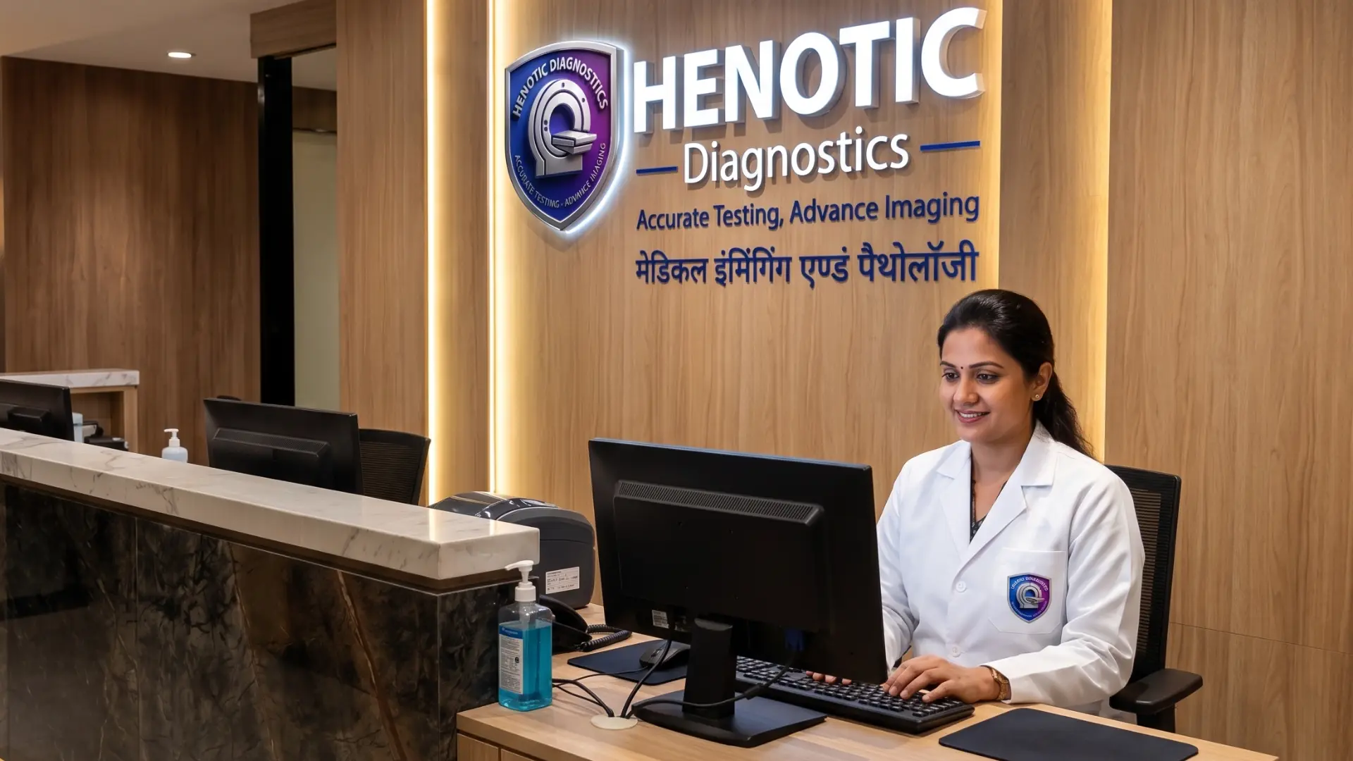
Specifically, efficient front-desk registration workflows minimize waiting times and ensure seamless clinical check-ins for all Radiology Services.
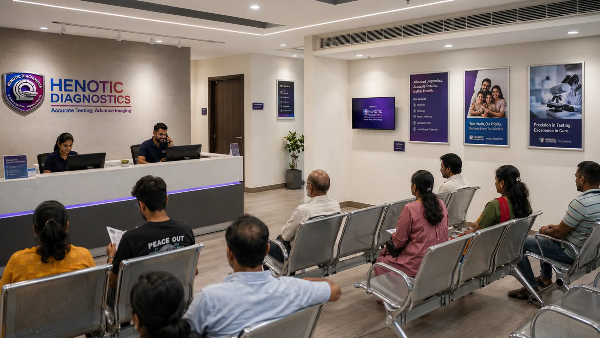
Additionally, spacious, climate-controlled reception lounges offer a relaxing atmosphere for patients and accompanying family members.
Furthermore, state-of-the-art imaging facilities integrate specialized patient care protocols to enhance convenience:
Diagnostic imaging interpretation at Henotic Diagnostics is led by our consultant radiologist.

MBBS MD (Radiodiagnosis)
Specifically, Dr. Pratibha Patil oversees the radiology diagnostic reporting workflow at Henotic Diagnostics. Furthermore, her clinical focus encompasses neuro-radiology MRI, CT angiography, pelvic sonography, and multi-modality cancer imaging.
Additionally, working alongside referring physicians, Dr. Patil ensures that every imaging study provides clear diagnostic findings to assist patient care.
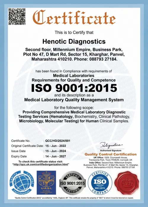
Fundamentally, diagnostic imaging quality and safety standards at Henotic Diagnostics align with national regulatory authorities and quality frameworks for Radiology Services.
.webp)
Radiation Safety & Diagnostic Equipment Quality Assurance

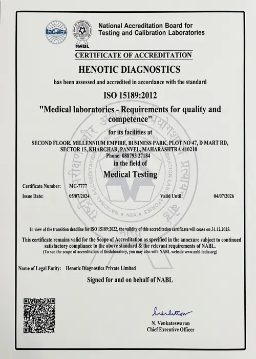
Laboratory Quality & Diagnostic Competency Accreditation

International Quality Management System Certified

International Society of Ultrasound in Obstetrics & Gynecology

Healthcare Facility Quality & Patient Safety Protocols

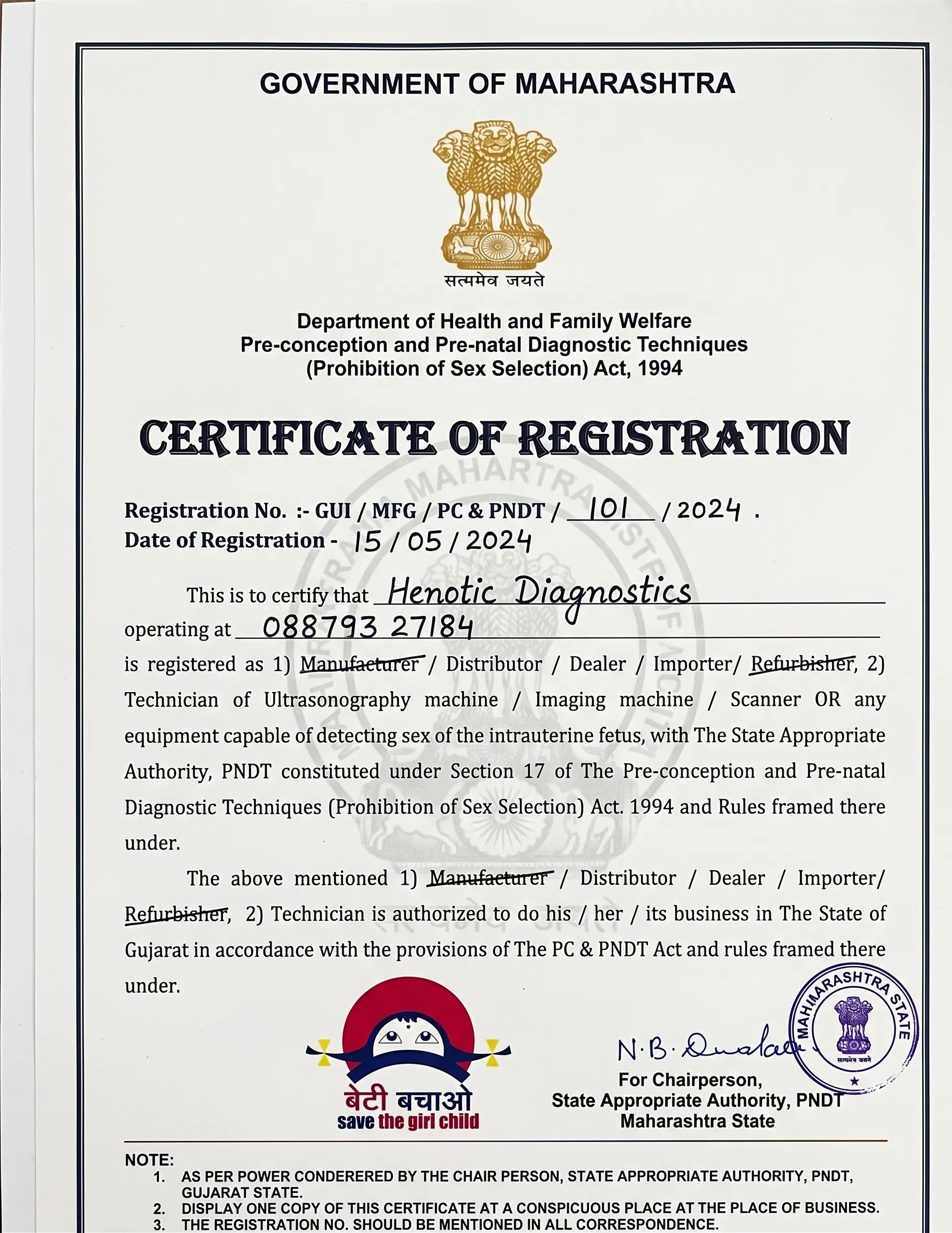
Full Compliance with Pre-Conception & Pre-Natal Diagnostic Techniques Act
Specifically, Henotic Diagnostics maintains complete transparent documentation and regulatory approvals for all Radiology Services:


Read patient experiences regarding communication, scheduling, and diagnostic reporting at Henotic Diagnostics.
“I underwent an MRI spine scan at Henotic Diagnostics. Furthermore, the MRI room was clean, the staff explained the procedure clearly, and the digital report was delivered to my doctor promptly.”
“My doctor requested an abdominal ultrasound and Doppler study. In addition, the sonographer was gentle, efficient, and ensured my comfort throughout the examination.”
“The CT scan procedure was fast and well-coordinated. Consequently, receiving digital report access on my phone saved me an extra trip to collect physical films.”
Henotic Diagnostics is located in Sector 15, Kharghar, offering scheduling support for all diagnostic imaging appointments.
Address: Second floor, Millennium Empire, Business Park, Plot No 47, D Mart Rd, Sector 15, Kharghar, Navi Mumbai, Panvel, Maharashtra 410210
Phone / Booking Helpline:
+918879327184
24 Hours Open

Specifically, radiology services encompass advanced diagnostic imaging techniques—including MRI, CT, ultrasound, Doppler, X-ray, mammography, PET-CT, DEXA, and nuclear medicine—used to visualize anatomical structures, assess physiological processes, and detect medical conditions accurately.
Primarily, a radiologist is a specialized medical doctor who interprets diagnostic imaging scans, provides expert diagnostic consultations to clinical physicians, guides image-guided procedures, and ensures radiation safety during imaging examinations.
For instance, the main modalities of radiology imaging are Magnetic Resonance Imaging (MRI), Computed Tomography (CT), Diagnostic Ultrasound & Sonography, Color Doppler Ultrasound, Digital Radiography (X-Ray), Digital Mammography, PET-CT Imaging, Nuclear Medicine, and Bone Mineral Density (DEXA) scans.
Undeniably, Magnetic Resonance Imaging (MRI) is an essential non-invasive subspecialty of diagnostic radiology services that utilizes strong magnetic fields and radiofrequency pulses to capture high-resolution cross-sectional soft tissue images without ionizing radiation.
Indeed, diagnostic ultrasound (sonography) and Doppler imaging are core components of radiology services that use high-frequency sound waves to visualize internal organs, vascular hemodynamics, and fetal development in real-time.
Consequently, Computed Tomography (CT) utilizes low-dose ionizing X-ray radiation combined with computer processing to reconstruct multiplanar cross-sectional images of internal body structures.
In contrast, MRI does not use ionizing radiation. Specifically, it relies entirely on radio waves and strong magnetic fields, making it safe for repeat structural soft tissue evaluation.
Whereas CT scans use X-rays to generate rapid images of bony structures, chest pathology, and acute hemorrhages, MRI uses magnetic fields to deliver detailed soft tissue contrast resolution for brain, spine, joint, and pelvic evaluations.
Fundamentally, radiology services provide accurate, non-invasive visualization of internal pathology, allowing early disease detection, precise treatment planning, staging of oncological conditions, and objective monitoring of clinical recovery.
Accordingly, an imaging test is selected based on clinical presentation, targeted anatomical organ system, diagnostic sensitivity, safety considerations (such as pregnancy or renal function), and referral guidelines from treating physicians.
Indeed, Typically, during an imaging exam, certified radiologic technologists position the patient on specialized imaging tables, guide breathing instructions if required, acquire diagnostic scans, and transmit images to radiologists for interpretation.
Additionally, patient preparation varies by test: abdominal ultrasounds may require 6 hours of fasting, pelvic ultrasounds require a full bladder, contrast CT/MRI scans require kidney function testing (creatinine), and MRI exams require removing metallic objects.
Ultimately, licensed consultant radiologists analyze imaging datasets, evaluate morphological and functional findings, correlate with clinical history, and generate formal diagnostic reports.
Get priority booking through our secure portal.