
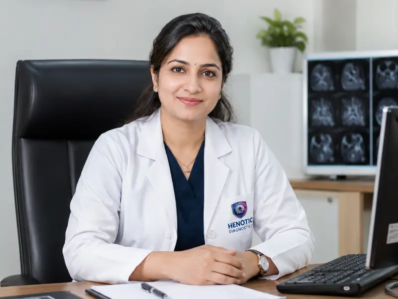
Dr Pratibha Patil
MBBS, MD (Radio-Diagnosis)
16+ Years Clinical ExperienceSpecialist in: 3D Digital Breast Tomosynthesis, Microcalcification Morphology, ACR BI-RADS Staging, Stereotactic Interventions, and Women’s Imaging
Connecting to our secure server
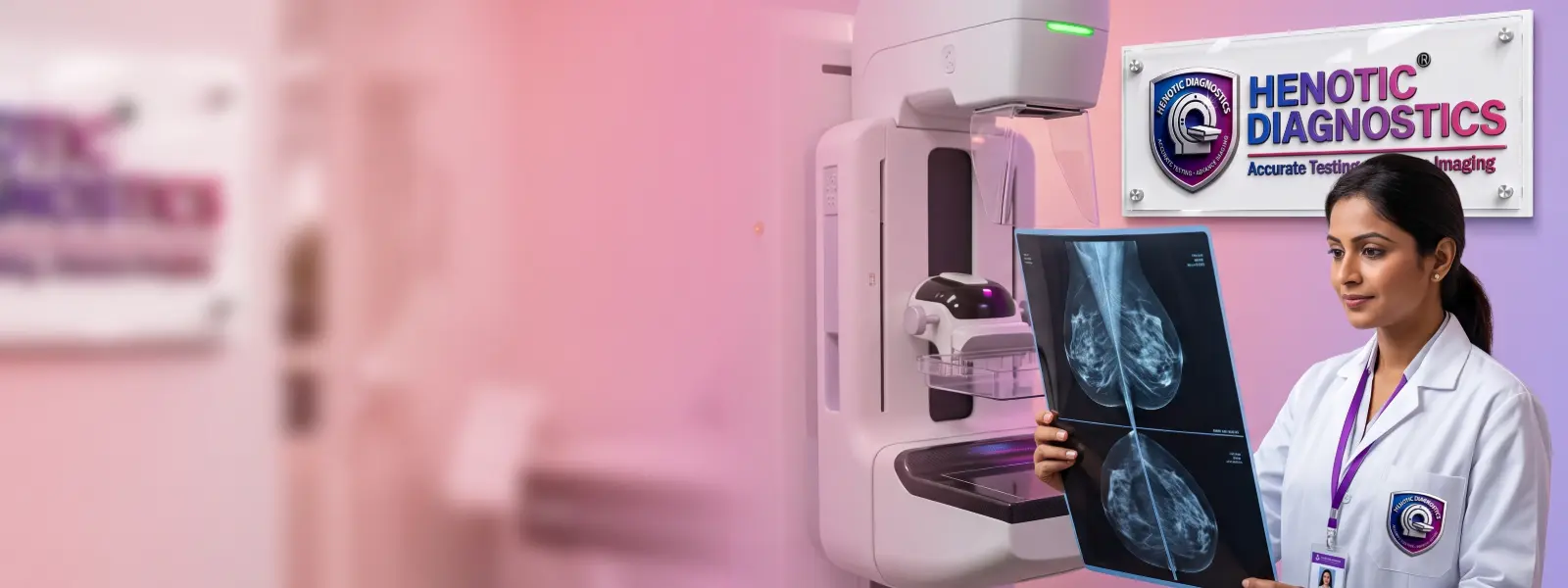
Browse our comprehensive library of diagnostic capabilities. We provide over 200 NABL accredited radiology scans and automated clinical pathology investigations.
Specifically, our advanced Digital Breast Tomosynthesis (DBT) captures high-resolution 1mm volumetric slices, eliminating tissue overlap to detect subtle lesions early with low radiation at Henotic Diagnostics, Kharghar.

Furthermore, the comparison table below summarises key technical parameters you need to understand prior to your Digital Breast Tomosynthesis appointment.
| Parameter | Clinical Specification |
|---|---|
| Service Modality | 3D Digital Breast Tomosynthesis (DBT) with Synthesized 2D Correlation |
| Imaging Mechanism | Motorized X-ray tube sweeps in an arc, capturing multiple thin 1mm projection slices |
| Primary Clinical Advantage | Eliminates tissue superposition to detect invasive tumours hidden in dense parenchyma |
| Radiation Standard | AERB-compliant ultra-low radiation dose comparable to conventional 2D mammography |
| Scan Acquisition Time | Rapid arc sweep — approximately 3.7 to 4 seconds of exposure per breast view |
| Compression Experience | Curved, ergonomic smart paddles reduce compression pressure by up to 30% |
| Reporting Classification | ACR BI-RADS Staging (Categories 0 through 6) by fellowship-trained radiologists |
| Turnaround Time | Same day — digital report with high-resolution image slices delivered in 2 to 4 hours |
| Specialist Supervision | Senior female radiologist review on calibrated multi-megapixel diagnostic monitors |
| Insurance Support | ✓ Accepted — Cashless TPA & Corporate Reimbursement Available |
| Diagnostic Facility | Henotic Diagnostics, Sector 15, Kharghar, Navi Mumbai (Open 24×7) |
| Direct Booking Desk | Call / WhatsApp: 088793 27184 |
Digital Breast Tomosynthesis is a revolutionary 3D mammographic screening and diagnostic modality that reconstructs a full volumetric dataset of breast architecture. Specifically, early detection using advanced volumetric mammography improves long-term clinical outcomes, as supported by the World Health Organization (WHO) Global Breast Cancer Initiative.
Specifically, during the examination, the X-ray tube glides smoothly in an arc over the breast, acquiring multiple low-dose projection images at distinct angles. Consequently, powerful reconstruction algorithms convert these exposures into a stack of 1mm thin cross-sectional slices. Indeed, radiologists can inspect the breast layer by layer, peeling away overlapping tissue that commonly conceals tumours in conventional planar imaging.
Additionally, at Henotic Diagnostics, we combine Digital Breast Tomosynthesis with high-resolution Bilateral Sono Mammography and Colour Doppler ultrasound. Consequently, this dual-modality synergy ensures comprehensive evaluation for dense breasts, microcalcifications, and architectural distortions.

Moreover, Digital Breast Tomosynthesis is universally recognized as the gold standard in diagnostic mammography. Furthermore, clinical evidence proves it resolves the fundamental limitations of traditional 2D imaging.
Furthermore, dense breast tissue masks tumours on 2D films because both appear radiopaque (white). Consequently, Digital Breast Tomosynthesis isolates lesions within 1mm slices, preventing healthy tissue from concealing suspicious micro-calcifications.
Moreover, patients with first-degree relatives or BRCA mutations benefit from annual 3D imaging, consistent with recommendations from the National Comprehensive Cancer Network (NCCN) Guidelines.
Additionally, annual 3D tomosynthesis screening for women aged 40 and above dramatically reduces interval cancers, endorsed by the American Cancer Society early detection recommendations.
Specifically, if you or your clinician identify a palpable lump, localized breast thickening, or discharge, 3D tomosynthesis provides precise margin characterization to guide clinical decisions.
Importantly, overlapping normal tissue on 2D mammograms often triggers anxious recalls and unnecessary biopsies. Digital Breast Tomosynthesis cuts recall rates by up to 30% by clarifying benign tissue overlap.
Furthermore, patients seeking thorough evaluations can bundle 3D tomosynthesis with complete women’s health checkup packages for holistic preventive wellness.
Conveniently, the table below demonstrates how 3D Digital Breast Tomosynthesis outperforms conventional 2D imaging, grounded in diagnostic evidence from the NHS Breast Screening Programme.
| Clinical Parameter | 3D Digital Breast Tomosynthesis | Conventional 2D Mammography | Sono Mammography (USG) | Breast MRI |
|---|---|---|---|---|
| Tissue Superposition | ✓ Eliminated (1mm Slices) | ✗ Severe Overlap | ✓ None (Real-time) | ✓ None |
| Invasive Cancer Detection | ✓ Up to 40% Higher | ✗ Standard Baseline | ✓ High in Dense Tissue | ✓ Highest Sensitivity |
| Dense Breast Accuracy | ✓ Outstanding | ✗ Significantly Reduced | ✓ Excellent | ✓ Excellent |
| Microcalcification Detection | ✓ Superior Sharpness | ✓ Good | ✗ Limited for Microcalcifications | ✗ Poor |
| False Recall Rate | ✓ 15%–30% Lower | ✗ Higher False Positives | ✓ Low with Doppler | ✗ Moderate False Recalls |
| Radiation Exposure | AERB Low-Dose Protocol | Low-Dose X-Ray | ✓ Zero Radiation | ✓ Zero Radiation |
| Compression Experience | ✓ Gentle Ergonomic Paddle | ✗ Rigid Compression | ✓ No Compression | ✓ None (Prone Coil) |
| Diagnostic Availability | ✓ 24×7 at Henotic | Standard Diagnostic | ✓ 24×7 at Henotic | Requires Prior Scheduling |
Specifically, Digital Breast Tomosynthesis allows radiologists to scroll through consecutive 1mm images of the breast, revealing micro-structures invisible on traditional planar films.
| Pathological Feature | Tomosynthesis Diagnostic Value |
|---|---|
| Clustered Microcalcifications | Analyzes morphology, branching patterns, and spatial distribution with sub-millimeter precision |
| Spiculated Masses | Uncovers fine radiating spicules of malignant tumours obscured by overlying fibrous tissue |
| Architectural Distortions | Identifies focal parenchymal tethering and subtle retractions indicative of early tubular carcinomas |
| Asymmetrical Densities | Resolves whether a focal density is a true underlying mass or harmless overlapping tissue |
| Circumscribed Lesions | Delineates sharp, smooth borders characteristic of benign fibroadenomas or simple cysts |
| Retroareolar Ductal Anatomy | Evaluates subareolar ductal dilatation, micro-masses, or ductal carcinoma in situ (DCIS) |
| Axillary Lymph Node Bases | Screens low axillary lymph nodes for cortical thickening, calcifications, or enlargement |
| Skin & Trabecular Thickening | Assesses cutaneous oedema or lymphatic engorgement associated with inflammatory disease |


Moreover, every scan at Henotic Diagnostics is conducted in an exclusive, private mammography suite by certified female technicians, adhering to strict AERB radiation safety standards.
Initially, our medical reception logs your symptoms and uploads previous 2D mammograms or ultrasound discs to the PACS workstation for comparative baseline analysis.
Next, a specialized female technologist reviews your family history, menopausal stage, prior biopsies, and hormone use to tailor the acquisition protocol.
Subsequently, the breast is gently positioned on the detector plate. Specifically, our curved ergonomic compression paddle applies customized, even pressure without acute pain.
Then, the X-ray tube sweeps in a swift 15-degree arc over the breast for approximately 3.7 seconds, capturing multiple micro-dose projection exposures.
Meanwhile, high-speed graphics processing units reconstruct raw exposures into a continuous stack of 1mm high-contrast cross-sectional slices.
Following 3D reconstruction, smart algorithms synthesize a 2D composite image (C-View), eliminating the need for an additional radiation-exposing 2D acquisition.
Afterward, our senior radiologists scroll through the volumetric slice stack on certified 5-megapixel medical displays, assessing every millimeter under BI-RADS guidelines.
Finally, your comprehensive 3D mammography report is delivered within 2 to 4 hours via WhatsApp and digital portal, complete with annotated slice captures.
Indeed, your Digital Breast Tomosynthesis examination is interpreted exclusively by senior consultant radiologists with fellowship accreditation in advanced breast imaging.
MBBS, MD (Radio-Diagnosis)
16+ Years Clinical ExperienceSpecialist in: 3D Digital Breast Tomosynthesis, Microcalcification Morphology, ACR BI-RADS Staging, Stereotactic Interventions, and Women’s Imaging

MBBS, DNB (Radio-Diagnosis)
11+ Years Clinical ExperienceSpecialist in: Breast Tomosynthesis Interpretation, Sono Mammography Correlation, USG-Guided FNAC, Core Needle Biopsy, and Vascular Sonology
Importantly, simple pre-examination preparation ensures crystal-clear image acquisition and avoids artifacts that could mimic microcalcifications.
| ✓ Recommended Preparations |
|---|
| Wear a comfortable two-piece outfit (skirt/trousers and top) for ease of changing |
| Bring previous 2D mammography films, ultrasound reports, and surgical summaries |
| Schedule the scan the week after your menstrual period when breasts are least tender |
| Notify our technician if you have breast implants, pacemakers, or are breastfeeding |
| Furthermore, carry your health insurance card for cashless billing desk verification |
| Arrive 15 minutes early to complete clinical questionnaire profiling comfortably |
| ✗ What to Avoid |
|---|
| Do not apply deodorants, antiperspirants, or body sprays to underarms or chest |
| Avoid using talcum powder, perfumes, or metallic-particle shimmer body lotions |
| Refrain from wearing neck jewellery or metallic ornaments during the acquisition |
| Do not postpone your appointment if you feel a palpable lump — urgent booking is vital |
| Therefore, avoid taking caffeine heavily on test morning if you suffer cyclic breast pain |

Specifically, Henotic Diagnostics operates state-of-the-art 3D tomosynthesis platforms featuring high-detective quantum efficiency (DQE) amorphous selenium detectors, adhering to diagnostic parameters outlined by the Radiological Society of North America (RSNA).
Furthermore, our platform incorporates synthetic 2D image reconstruction. Specifically, this cutting-edge mathematical algorithm generates a crystal-clear 2D mammographic view straight from the 3D tomosynthesis slice dataset, eliminating the need for an additional 2D exposure and keeping patient dose well below AERB regulatory limits.
Additionally, ergonomic curved paddles adapt to the natural contour of the breast, distributing compression pressure uniformly across the gland. Consequently, discomfort is significantly lessened while achieving optimal tissue immobilization.
Moreover, all examinations are evaluated under the rigorous American College of Radiology (ACR) BI-RADS Atlas on Barco 5-megapixel medical-grade diagnostic monitors.
Moreover, Henotic Diagnostics adheres to the highest national and international radiation safety and quality benchmarks for Digital Breast Tomosynthesis.
 AERB Approved
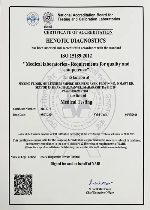 NABL Accredited
 CAP Certified
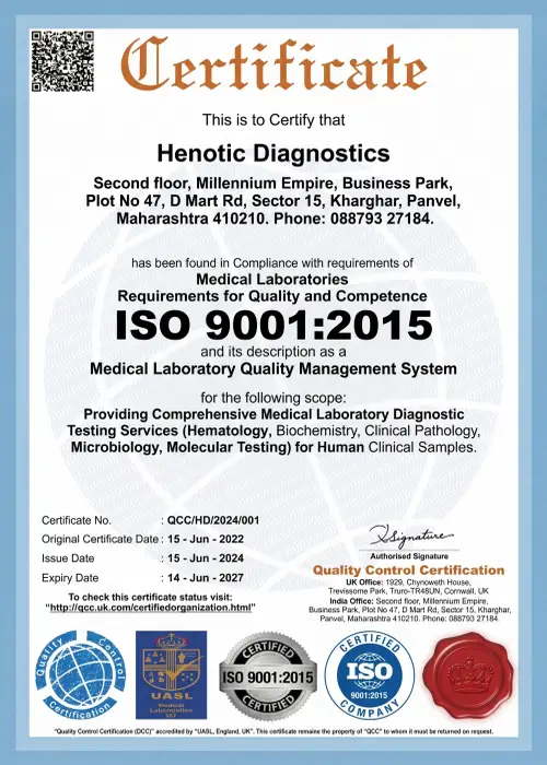 ISO 9001:2015
 ISUOG Member
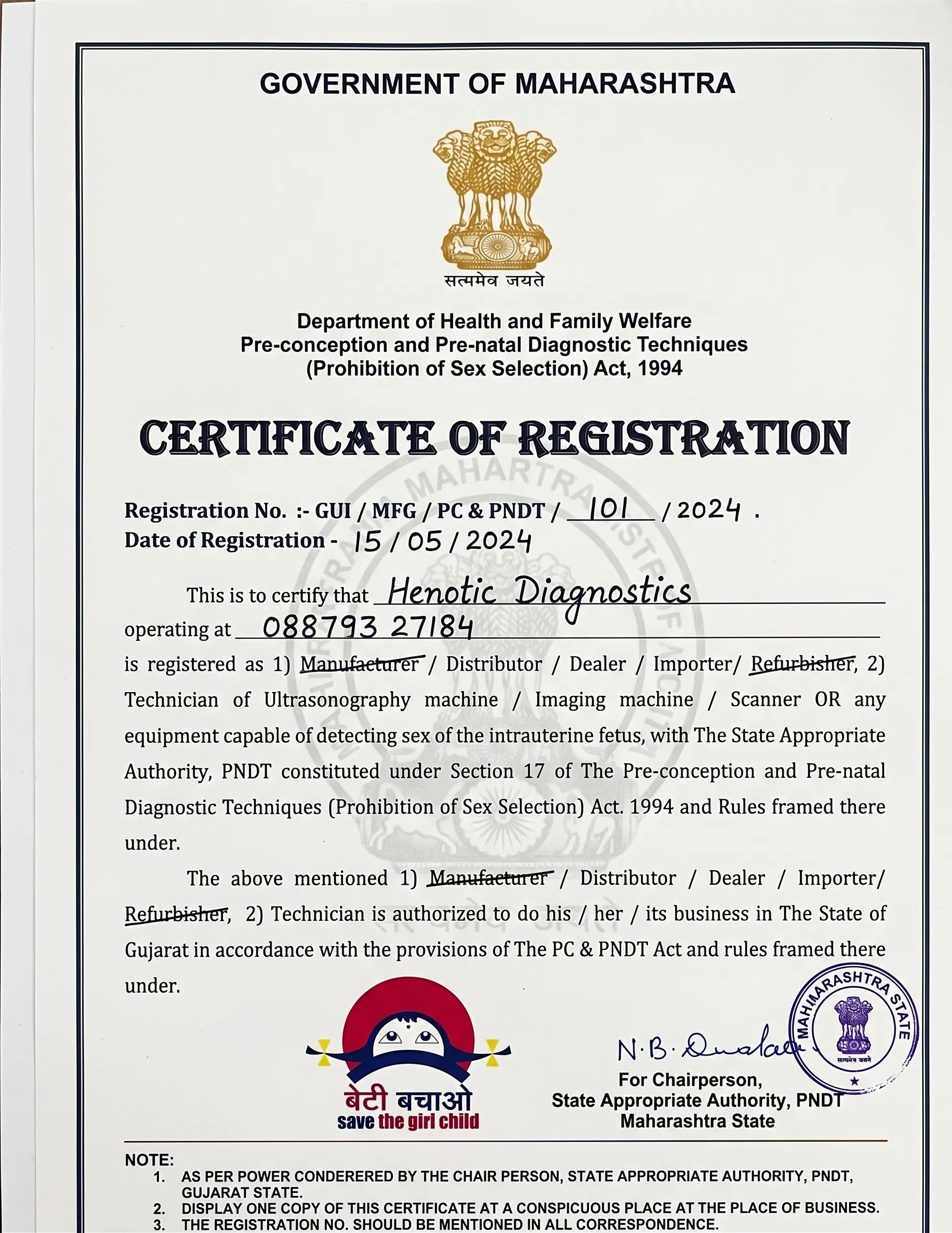 PCPNDT Registered
Additionally, our verified Google patient reviews reflect the compassionate, dignified care and diagnostic precision delivered across every 3D mammography consultation.
“Indeed, I was terrified of mammograms after a painful experience elsewhere. Conveniently, at Henotic Diagnostics, the curved paddle was surprisingly gentle. The 3D Digital Breast Tomosynthesis scan was over in seconds, and Dr. Patil explained my normal BI-RADS 1 report directly on the screen.”
“Moreover, having dense breast tissue, my previous 2D mammogram missed a tiny fibroadenoma that caused me endless anxiety. In fact, the 3D tomosynthesis at Henotic clearly differentiated the benign nodule from surrounding glandular tissue. Exceptional service and prompt WhatsApp reports.”
“Transparent billing, no hidden fees, and seamless cashless processing with my insurance provider. Consequently, I will never go back to older 2D mammography. Henotic Diagnostics is clearly the premier centre in Navi Mumbai.”
Specifically, every 3D mammography report at Henotic Diagnostics is classified according to the American College of Radiology BI-RADS system for universal clinical clarity.
| BI-RADS Category | Digital Breast Tomosynthesis Finding | Recommended Clinical Protocol | Malignancy Likelihood |
|---|---|---|---|
| Category 0 | Incomplete — requires additional spot-compression or targeted ultrasound | Supplementary bilateral sono mammography or MRI correlation | Undetermined |
| Category 1 | Negative — symmetrical glandular tissue, no microcalcifications or masses | Routine annual Digital Breast Tomosynthesis screening | Essentially 0% |
| Category 2 | Benign finding — documented secretory calcifications, typical fibroadenoma | Routine annual screening follow-up | 0% |
| Category 3 | Probably benign — well-circumscribed oval mass or focal asymmetry | Short-interval 6-month unilateral repeat tomosynthesis | <2% |
| Category 4 | Suspicious abnormality — irregular margins or clustered microcalcifications | USG-guided FNAC or stereotactic core needle biopsy | 2%–95% |
| Category 5 | Highly suggestive of malignancy — spiculated mass with pleomorphic calcifications | Urgent tissue biopsy and surgical oncology consultation | >95% |
| Category 6 | Known biopsy-proven malignancy — staging and neoadjuvant response tracking | Surgical excision or oncology management protocol | 100% (Confirmed) |
Conveniently, Henotic Diagnostics is empanelled with leading health insurance providers and TPAs for seamless Digital Breast Tomosynthesis claims processing.
| Insurance Provider | Coverage for 3D Tomosynthesis | Documentation Required |
|---|---|---|
| Star Health | ✓ Covered under OPD / Diagnostic Benefits | Insurance card + Photo ID + Doctor prescription |
| HDFC ERGO | ✓ Covered under comprehensive health plans | Policy number + Treating physician referral |
| New India Assurance | ✓ Covered for diagnostic indications | Policy certificate + Clinical notes + ID proof |
| Niva Bupa | ✓ Covered with OPD diagnostic rider | Digital health card + Prescription |
| Medi Assist TPA | ✓ Cashless pre-authorization supported | TPA membership card + Referral |
| Ayushman Bharat / PMJAY | ✓ Covered for eligible empanelled categories | PMJAY e-card + Aadhaar verification |
| Corporate Group Health | ✓ Most corporate executive health packages | Employee corporate ID + Policy card |
Importantly, call our insurance desk at 088793 27184 to confirm your cashless eligibility prior to your scan.
Importantly, the following clinical questions address common patient inquiries regarding 3D mammography and Digital Breast Tomosynthesis.
Specifically, Digital Breast Tomosynthesis is a 3D mammography technique where an X-ray tube arcs over the breast to capture multi-angle exposures. These are reconstructed into 1mm thin slices, eliminating overlapping tissue and dramatically improving early cancer visualization.
Furthermore, all women aged 40 and older, women of any age with dense breast tissue, or individuals with a family history of breast disease should choose 3D tomosynthesis, in accordance with CDC Breast Cancer Screening guidance.
Indeed, at Henotic Diagnostics, our tomosynthesis unit employs ergonomic curved paddles that adjust to breast curvature, preventing pinched skin. Compression duration is brief — taking only 3.7 to 4 seconds of exposure per projection view.
In fact, traditional 2D mammography produces a flat projection where dense glandular tissue often hides malignant masses. Digital Breast Tomosynthesis creates multi-layered cross-sections, increasing invasive cancer detection by up to 40% while cutting false alarms.
Specifically, modern tomosynthesis systems utilize advanced synthetic 2D algorithms (C-View), which eliminate the need for a separate 2D exposure. Consequently, the radiation dose is extremely low and fully compliant with AERB safety thresholds.
Conveniently, at Henotic Diagnostics, your Digital Breast Tomosynthesis study is reviewed on high-resolution 5MP monitors by senior female radiologists, with complete BI-RADS reports delivered within 2 to 4 hours via WhatsApp and digital portal.
Furthermore, Henotic Diagnostics offers a comprehensive spectrum of NABL and AERB-accredited imaging and pathology investigations, operating 24 hours a day, 7 days a week.
Conveniently, Henotic Diagnostics is located at Sector 15, Kharghar, Navi Mumbai — easily accessible from Belapur, Panvel, Vashi, Nerul, and Kamothe for 3D mammography appointments.
Simply fill in the details below to reserve your priority 3D mammography appointment at Henotic Diagnostics. Alternatively, you can call or WhatsApp 088793 27184 for direct coordination.
Get priority booking through our secure portal.