
Loading Diagnostics...
Connecting to our secure server
Connecting to our secure server
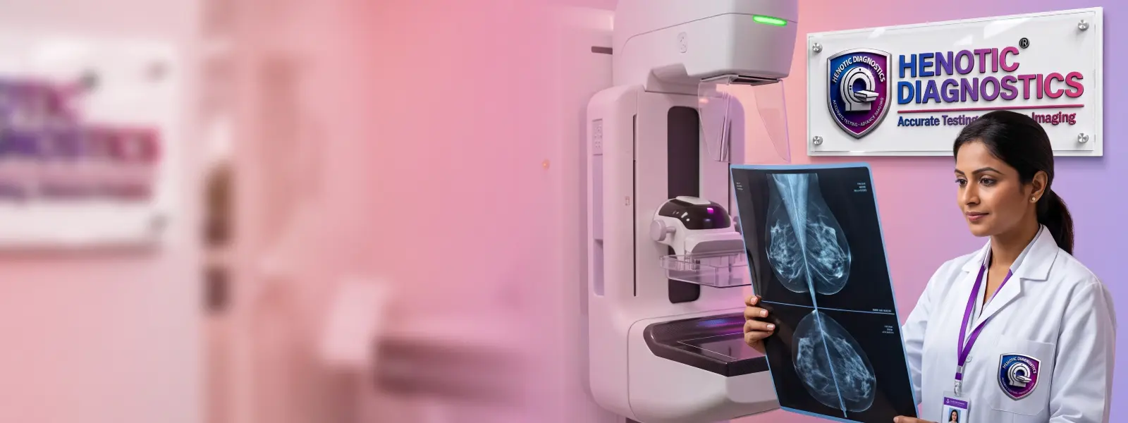
Browse our comprehensive library of diagnostic capabilities. We provide over 200 NABL accredited radiology scans and automated clinical pathology investigations.
Accurate, radiation-free, high-resolution breast ultrasound interpreted by certified senior consultant radiologists.

Key parameters regarding examination preparation, technology, and report delivery.
| Service Information | Details |
|---|---|
| Examination | Sono Mammography / Breast Ultrasound |
| Imaging method | Ultrasound |
| Ionizing radiation | No |
| Fasting | Not required |
| Preparation | Minimal |
| Typical examination duration | Approximately 15–30 minutes |
| Patient experience | Generally well tolerated |
| Interpretation | Radiologist reporting |
| Digital reports | Yes |
| WhatsApp report delivery | Yes |
| Email report delivery | Yes |
| Hardcopy report | Available at centre on request |
| Centre availability | Open 24 hours |
| Appointment | Prior booking recommended |
Consultant radiologists specializing in Women’s Imaging and ultrasound diagnostics.

16+ years experience; Specialist in Women’s Imaging & BI-RADS.

11+ years experience; Expert in Breast Ultrasound & USG-Guided FNAC.
Verified diagnostic compliance and quality accreditation standards.
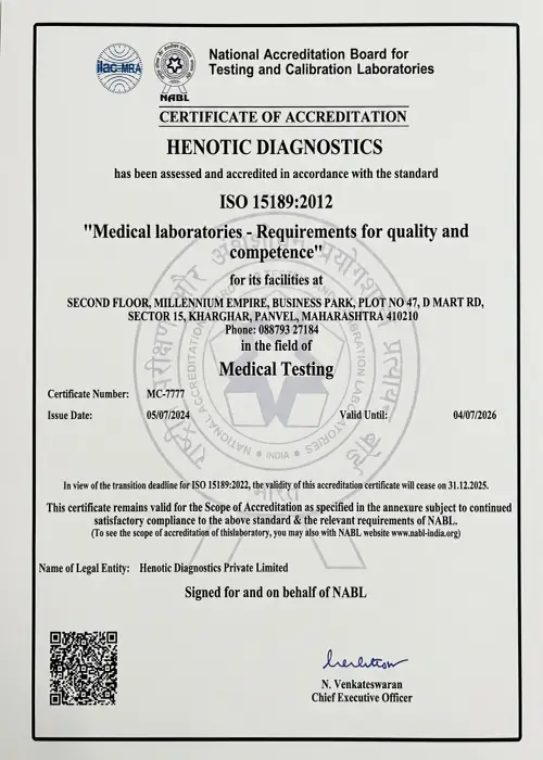 NABL
NABL
 CAP
CAP
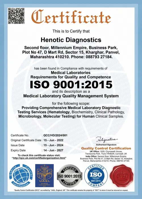 ISO:-9001:2015
 AERB
AERB
 ISUOG
ISUOG
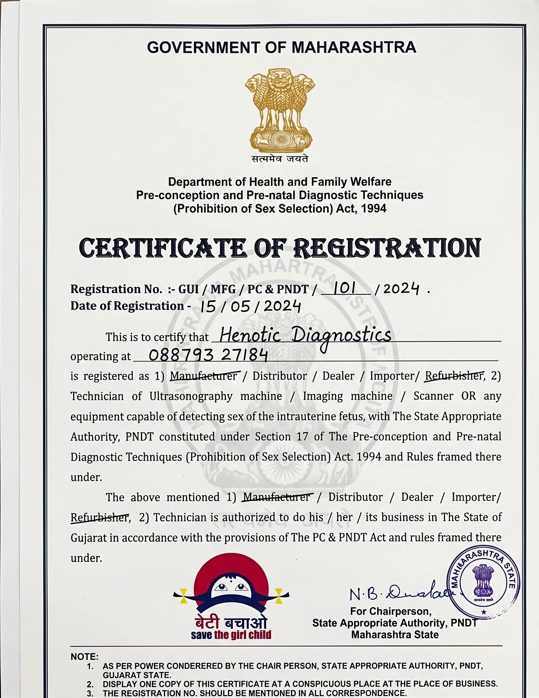 PCPNDT
Sono Mammography is an ultrasound examination specifically used to assess breast tissue.
During the examination, high-frequency sound waves are used to generate real-time images of the breast. A radiologist evaluates the images to assess the characteristics of breast tissue and any areas that require further attention.
Sono Mammography can help evaluate:
Unlike X-ray mammography, ultrasound does not use ionizing radiation.

Clinical indications for focused breast screening and diagnostic ultrasound evaluations.
Sono Mammography may be recommended when a clinician needs additional information about a breast symptom, palpable finding or imaging abnormality.
Common clinical indications include:
The appropriate breast-imaging examination depends on the patient’s age, symptoms, medical history, risk factors and clinical question.
Understanding the clinical purpose and modalities of each procedure.
| Sono Mammography | Mammography |
|---|---|
| Uses ultrasound waves | Uses low-dose X-rays |
| Does not use ionizing radiation | Uses ionizing radiation |
| Produces ultrasound images of breast tissue | Produces X-ray images of breast tissue |
| Useful for targeted evaluation of breast findings | Important breast imaging modality for screening and diagnostic evaluation |
| Can help distinguish cystic and solid lesions | Provides information about breast tissue and certain mammographic findings |
| May be used as an additional examination | May be recommended depending on age, risk and clinical circumstances |
Important: Sono Mammography does not automatically replace mammography. A doctor may recommend one examination or a combination of examinations depending on the clinical situation.
Characterisation of breast lumps, cysts, and architectural changes.
A breast lump can have several possible causes, including benign conditions.
Ultrasound can help the radiologist evaluate characteristics such as:
Dense Breasts: Breast density can affect the appearance of breast tissue on mammography. Ultrasound may be used as an additional imaging examination in selected patients when clinically appropriate.
During Pregnancy: Ultrasound does not use ionizing radiation and may be used when breast imaging is clinically required during pregnancy. If you are pregnant or think you may be pregnant, inform the radiology team before the examination.
| Ultrasound Assessment | Clinical Information |
|---|---|
| Cystic appearance | Helps identify fluid-containing lesions |
| Solid appearance | Requires assessment of morphology and other characteristics |
| Lesion margins | Contributes to imaging characterisation |
| Shape | Helps describe the imaging appearance |
| Internal features | Provides additional diagnostic information |
| Size | Allows measurement and documentation |
| Location | Identifies the anatomical site |
The imaging appearance is interpreted by the reporting radiologist and should be considered together with the clinical history.
Structured clinical workflow ensuring patient comfort, dignity, and accuracy.
Guidelines to prepare for your breast screening appointment.
| Requirement | Guidance |
|---|---|
| Fasting | Not required (eat and drink water normally) |
| Pain / Discomfort | Generally well tolerated; mild probe pressure may be felt on tender areas |
| Prescription | Bring your doctor’s prescription if available |
| Previous reports | Bring prior breast imaging reports, films, or CD/DVDs for radiologist comparison |
| Medical history | Inform the team about relevant personal/family breast history or pregnancy |
| Clothing | Comfortable two-piece attire is recommended |

High-quality breast ultrasound depends on appropriate equipment, suitable imaging protocols, accurate image acquisition and professional interpretation.
| Doppler Capability | High-Resolution Colour and Power Doppler |
| Radiation Exposure | None (Non-ionizing acoustic waves) |
| Center Availability | Open 24 Hours |
| Report Channels | WhatsApp, Email & Hardcopy |
Real feedback from verified patients at Henotic Diagnostics.
“I was extremely anxious after finding a lump. The female technician and doctor were gentle, explained everything on the screen, and delivered my report on WhatsApp within 2 hours. Highly recommended.”
“Very clean center with polite staff. No long waiting lines. Dr. Sharma reviewed my previous mammogram side-by-side with the sonomammography to confirm it was a benign cyst. Pure peace of mind.”
“Transparent pricing with no hidden charges. Claimed my OPD insurance smoothly with their structured receipts.”
Patient review source: Google Business Reviews
Answers to common clinical questions regarding breast screening.
Sono Mammography is an ultrasound examination of the breast that uses high-frequency sound waves to produce images of breast tissue and evaluate abnormalities such as lumps, cysts and masses.
No. Fasting is not required for routine Sono Mammography.
No. Ultrasound does not use ionizing radiation.
A typical Sono Mammography examination takes approximately 15–30 minutes, although the duration can vary depending on the examination and clinical findings.
It is generally well tolerated. Some patients may experience mild pressure while the ultrasound transducer is moved over the breast.
It can help evaluate breast lumps, cysts, solid masses, focal abnormalities and other clinically relevant breast findings.
No. Sono Mammography uses ultrasound, while mammography uses X-rays. The two examinations have different clinical roles and can sometimes be used together.
Ultrasound does not use ionizing radiation and may be used when breast imaging is clinically indicated during pregnancy. The appropriate examination should be determined by a healthcare professional.
Diagnostic imaging facility and appointment booking details.
Phone: 088793 27184
Opening Hours: Open 24 Hours
Get priority booking through our secure portal.