Loading Diagnostics...
Connecting to our secure server
Connecting to our secure server
Browse our comprehensive library of diagnostic capabilities. We provide over 200 NABL accredited radiology scans and automated clinical pathology investigations.
Specifically, a Radiology Center is an advanced medical imaging center dedicated to obtaining visual representations of the body’s internal anatomy, organ systems, micro-vascular blood flow, and physiological processes using non-invasive energy forms. Consequently, radiology centers play an essential role in modern healthcare by providing objective anatomical data needed by referring clinicians to diagnose diseases, guide targeted interventional procedures, evaluate traumatic injuries, and monitor therapeutic responses across oncology, neurology, cardiology, orthopedics, and gastroenterology.
Specifically, diagnostic radiology uses sub-specialty imaging techniques including advanced MRI scan protocols to capture anatomical cross-sections, cellular metabolic uptake, and real-time movement within human body structures for comprehensive clinical diagnostic evaluation.
Additionally, different modalities utilize distinct physical principles: strong magnetic fields, collimated X-ray beams in CT scan procedures, high-frequency acoustic waves, and radiotracers in PET-CT imaging.
For instance, scans evaluate cerebral white matter, spinal nerve roots, cardiac muscle perfusion, pulmonary parenchyma, solid abdominal organs via ultrasound and sonography, peripheral blood vessels, musculoskeletal cartilage, and skeletal bone density.
In contrast, radiology captures spatial structure, tissue density, and vascular perfusion non-invasively inside the living body using Color Doppler study modalities, while clinical laboratory analysis evaluates bio-fluids under microscopy.
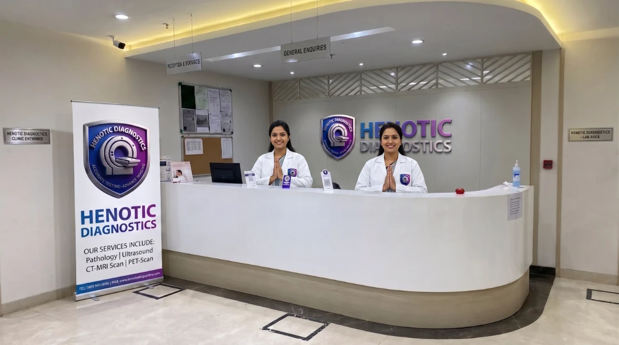
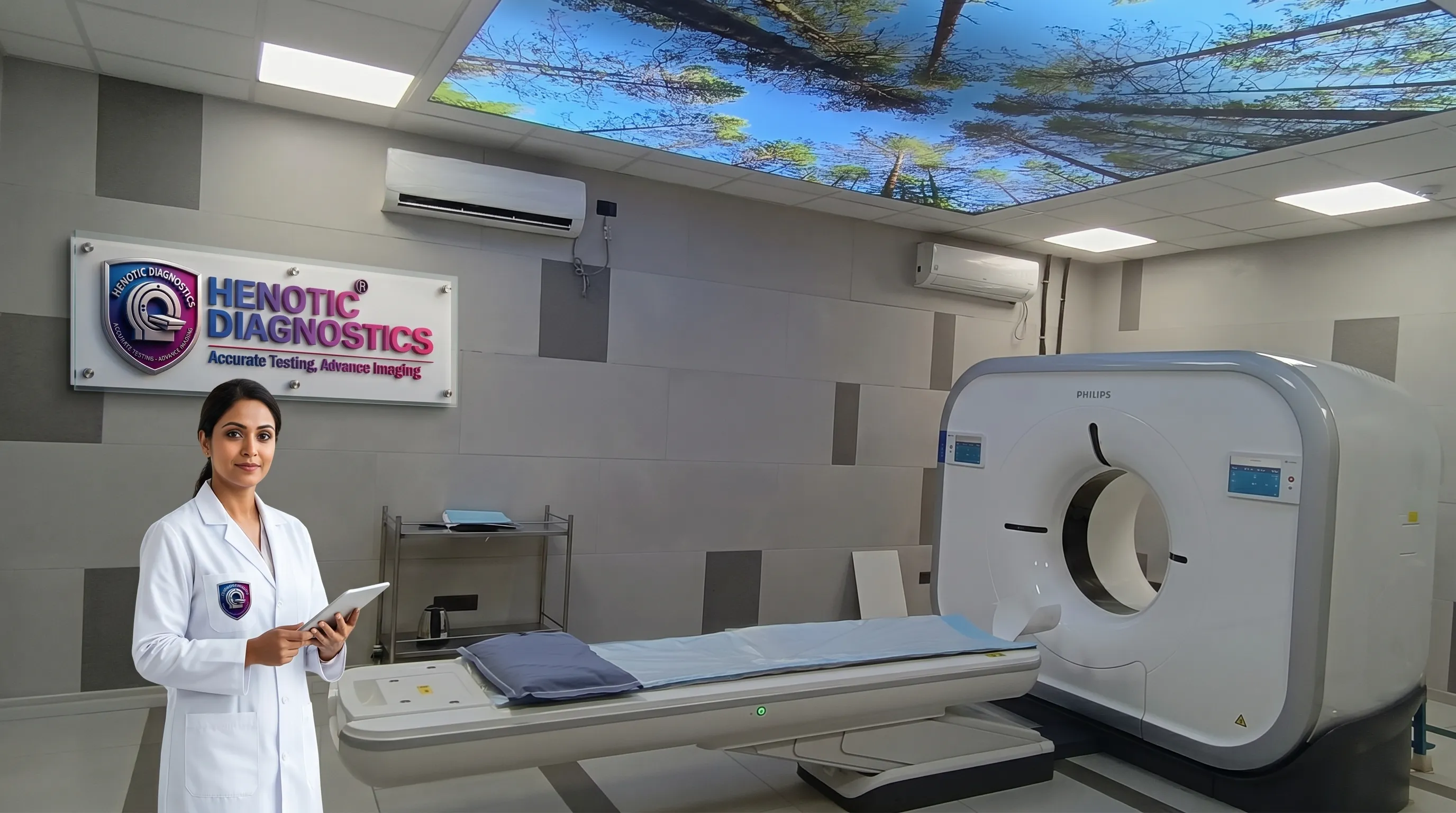
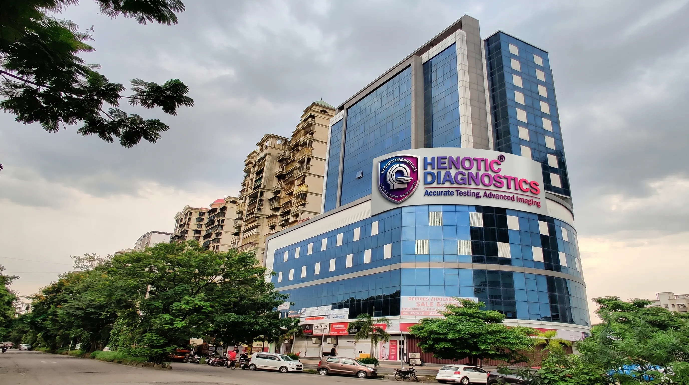
Furthermore, Henotic Diagnostics Outpatient Radiology Center & Diagnostic Imaging Suite Environment
Specifically, Henotic Diagnostics Radiology Center provides hospital-grade diagnostic imaging across 12 major specialized modalities, supported by calibrated equipment and sub-specialty radiologist reporting.
Specifically, high-field Magnetic Resonance Imaging provides non-ionizing anatomical and functional soft-tissue visualization with sub-millimeter spatial resolution at our facility.
For instance, brain neoplasms, stroke evaluation, spinal cord compression, joint cartilage tears, liver/pelvis mass characterization, vascular angiography, and cardiac viability.
Additionally, ultra-fast 128-slice Computed Tomography offers rapid multi-planar cross-sectional diagnostic reconstruction and 3D angiography.
For example, acute head trauma, interstitial lung disease, renal calculi, pulmonary embolism, coronary artery stenosis, complex bone fractures, and abdominal pathology.
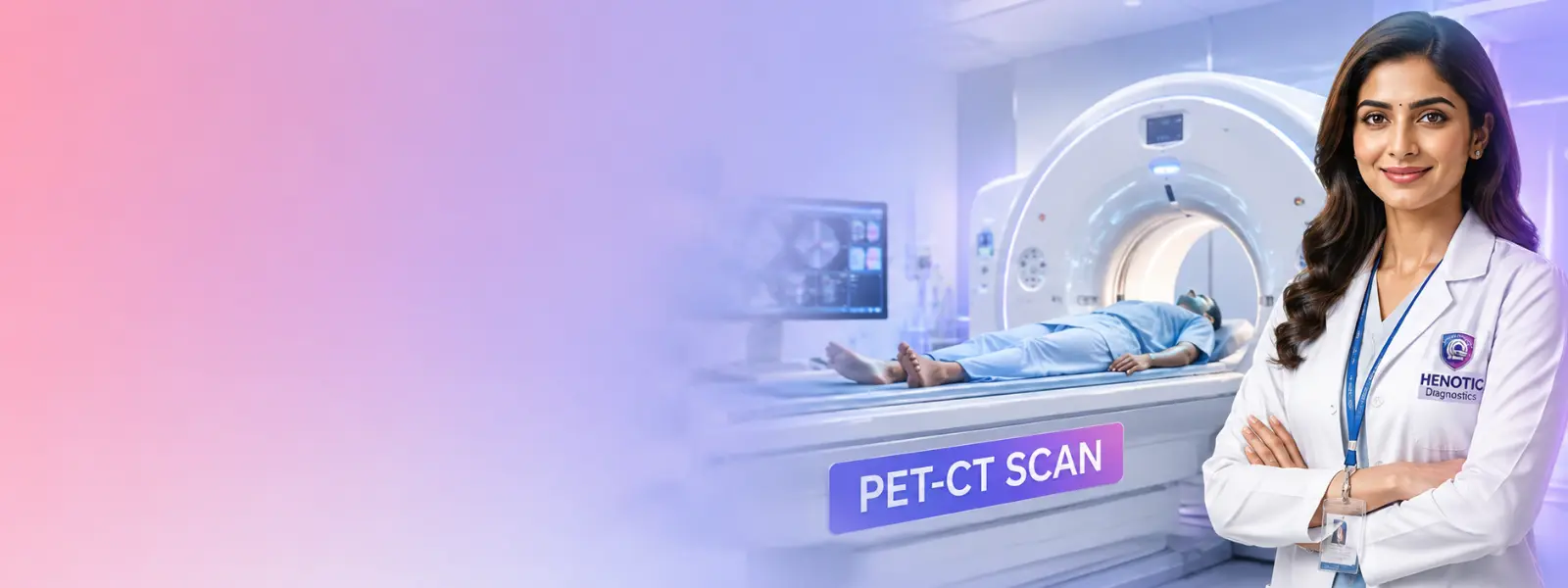
Furthermore, hybrid Positron Emission Tomography merged with CT maps cellular metabolic activity, tumor staging, and tracer perfusion, including specialized PSMA PET-CT imaging protocols.
Consequently, it evaluates oncology staging, treatment response, recurrence, prostate cancer (PSMA), neuroendocrine tumors (DOTA), and myocardial viability.
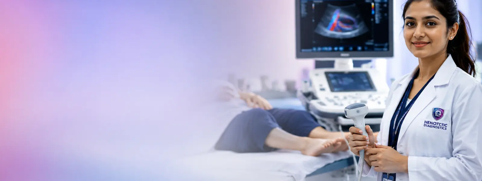
Moreover, high-resolution acoustic sonography uses multi-frequency transducers for real-time organ and soft-tissue evaluation.
As a result, ultrasound detects gallstones, fatty liver, renal tract calculi, thyroid nodules, pelvic pathology, scrotal pain, and guided tissue FNAC.
In addition, hemodynamic vascular sonography quantifies blood flow velocity, arterial narrowing, and venous thrombi using Carotid Doppler evaluation and limb vascular studies.
Specifically, it evaluates carotid artery stenosis, deep vein thrombosis (DVT), peripheral arterial disease (PAD), and fetal-placental blood flow.
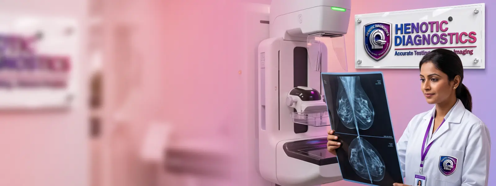
Similarly, low-dose digital mammography and 3D tomosynthesis enable early microcalcification detection.
Therefore, mammography provides routine breast screening, palpable lump investigation, and architectural distortion assessment.
Additionally, specialized obstetric ultrasound protocols track gestational development and fetal anatomical integrity.
For instance, pregnancy dating, nuchal translucency risk screening, detailed anomaly assessment, and growth monitoring.
Furthermore, advanced cardiovascular imaging evaluates coronary artery patency, valvular function, and cardiac muscle viability using 2D echocardiography studies and stress echo testing.
Consequently, cardiac scans detect coronary artery disease, heart valve disorders, myocardial scarring, and ejection fraction changes.
Specifically, real-time ultrasound or CT-guided minimally invasive needle aspiration and core biopsy procedures are performed by expert radiologists.
As a result, guided procedures obtain targeted tissue sampling of thyroid nodules, breast masses, liver lesions, and lymph nodes.
Additionally, non-invasive transient elastography delivers FibroScan liver assessment to measure stiffness and controlled attenuation parameter (CAP) for hepatic steatosis.
Specifically, it evaluates non-alcoholic fatty liver disease (NAFLD), chronic hepatitis fibrosis, and cirrhosis risk staging.
Moreover, Dual-Energy X-ray Absorptiometry provides precise DEXA bone density scan evaluations, calculating T-scores and Z-scores for osteoporosis staging.
Consequently, DEXA provides postmenopausal osteoporosis screening, fracture risk prediction, and body composition assessment.
Overall, high-frequency digital X-ray and dynamic fluoroscopy capture high-contrast skeletal and chest radiography with minimal radiation dose.
For example, bone fracture detection, pulmonary infection evaluation, osteoarthritis assessment, and pre-operative chest clearance.
Specifically, different medical symptoms require specific physical imaging modalities based on soft-tissue contrast, bone density, spatial resolution, and radiation factors.
| Clinical Area / Indication | Commonly Considered Diagnostic Imaging | Primary Diagnostic Objective |
|---|---|---|
| First, Brain & Neurological Indications | Specifically, MRI Brain examination (3T), CT Brain scan, MR Angiography | Consequently, detect stroke, tumors, aneurysms, demyelination, and acute hemorrhage. |
| Second, Spine & Spinal Cord Indications | Additionally, MRI Spine examination, Whole Spine MRI, CT Spine | Therefore, evaluate disc herniation, spinal stenosis, nerve compression, and trauma. |
| Third, Joints & Soft Tissue Indications | Furthermore, Joint MRI scan, Musculoskeletal Ultrasound | As a result, assess ligament tears, meniscus lesions, tendonitis, and soft-tissue masses. |
| Fourth, Chest & Lungs Indications | For example, HRCT Chest scan, Low-Dose Chest CT, Chest Radiography | Specifically, evaluate interstitial lung disease (ILD), nodules, pneumonia, and fibrosis. |
| Fifth, Abdomen & Viscera Indications | Moreover, Abdominal ultrasound, Abdomen CT scan, MRCP, MRI Abdomen | Consequently, diagnose gallstones, liver pathology, pancreatitis, appendicitis, and renal stones. |
| Clinical Area / Indication | Commonly Considered Diagnostic Imaging | Primary Diagnostic Objective |
|---|---|---|
| Sixth, Blood Vessels & Circulation Indications | In addition, Color Doppler study, CT Angiography, MR Angiography | Specifically, detect deep vein thrombosis (DVT), arterial stenosis, varicose veins, and aneurysm. |
| Seventh, Breast Health Indications | Similarly, Digital mammography, Sonomammography, Breast MRI | Therefore, screen microcalcifications, masses, architectural distortion, and dense tissue. |
| Eighth, Pregnancy & Fetal Care Indications | Additionally, Obstetric ultrasound, Level 2 Anomaly Scan, Fetal Doppler | For instance, monitor fetal growth, anatomical development, placental site, and cord flow. |
| Ninth, Cancer Evaluation Indications | Furthermore, PET-CT oncology imaging (FDG/PSMA), Contrast CT, MRI | Consequently, stage primary tumors, detect distant metastases, and evaluate treatment response. |
| Finally, Bone Density Indications | Overall, DEXA bone density scan (Lumbar Spine & Femur) | Specifically, quantify bone mineral density (BMD) and diagnose osteopenia/osteoporosis. |
Ultimately, accurate medical diagnosis relies equally on calibrated technological infrastructure, customized exam protocol selection, meticulous patient preparation, and expert sub-specialty radiologist interpretation.
Specifically, tailoring slice thickness, pulse sequences, contrast timing, and field-of-view in CT scan protocols answers specific clinical questions requested by referring physicians.
Furthermore, high magnetic field strength (3.0 Tesla) and sub-millimeter CT detectors preserve subtle structural details without motion artifacts or blurring.
Consequently, MRI scan interpretations are performed by board-certified radiologists with specific expertise in neuroradiology, musculoskeletal, cardiac, and abdominal imaging.
Additionally, high-speed DICOM PACS digital archiving enables instant comparison with historical imaging and seamless sharing with treating specialists.
In addition, board-Certified Radiologist & Sub-Specialty Diagnostic Imaging Analysis Workstation
Specifically, our 3T wide-bore MRI system operates at 3.0 Tesla magnetic field strength with silent scan acoustic reduction software and a 70cm wide bore, significantly reducing claustrophobia while capturing high-definition neuro, joint, and pelvic imaging.
Additionally, featuring a 128-slice CT platform with sub-millimeter physical detector channels permits sub-second whole-body rotation, allowing ultra-low-dose HRCT chest scans and high-frame-rate 3D coronary angiography.
Furthermore, integrating a hybrid PET-CT scanner with time-of-flight crystals provides precise co-registration of tumor metabolic activity with anatomical landmarks.
Moreover, high-end acoustic architecture in our Voluson 4D ultrasound supporting matrix array transducers enables multi-planar fetal echocardiography, high-frequency vascular Doppler, and elastography.
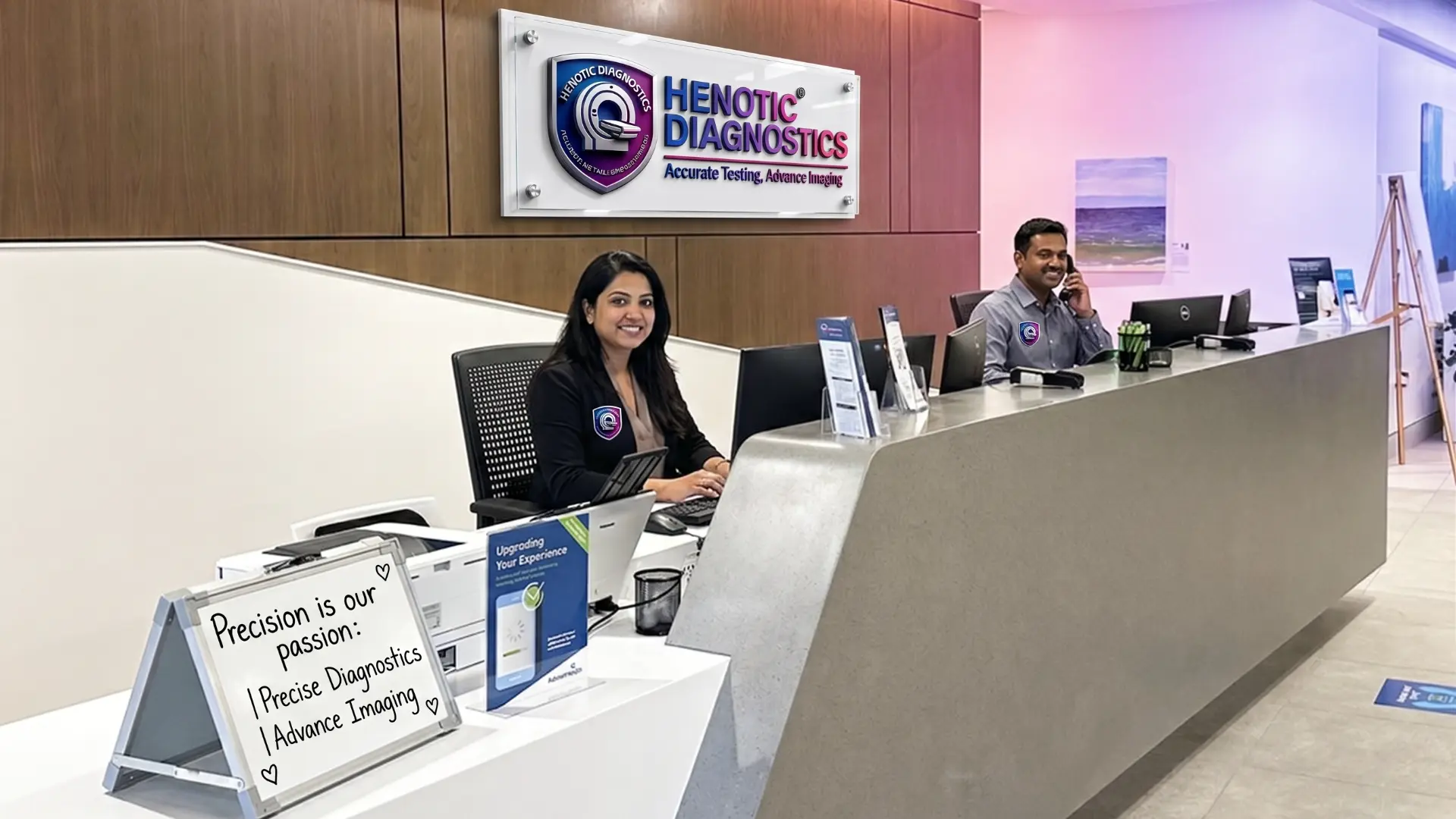
Additionally, NABL & AERB Accredited Patient Reception & Quality Control Desk
Specifically, our Radiology Center leverages enterprise Picture Archiving and Communication Systems (PACS) integrated with high-speed tele-radiology infrastructure to provide instant DICOM scan access and sub-specialty second opinion interpretations.
First, referring physicians can view high-resolution 3D DICOM cross-sections on desktop or mobile browsers without downloading specialized software.
Second, patients receive a secure digital portal link and QR code for instant report download and image sharing with consulting specialists.
Finally, all diagnostic scans are securely archived in redundant cloud storage for long-term comparative evaluation across future hospital admissions.
Specifically, Henotic Diagnostics Radiology Center provides instant digital access to signed diagnostic reports, high-resolution DICOM scan images, and comparative historical studies.
First, scan the secure QR code printed on your registration receipt using any smartphone camera to open your personal diagnostic portal dashboard immediately.
Second, as soon as your radiologist signs off your report, a secure encrypted link is sent automatically to your registered WhatsApp and mobile phone.
Third, download official digitally signed diagnostic PDF reports containing high-resolution key image slices, quantitative tables, and radiologist impressions.
Finally, generate encrypted temporary viewing links to share full DICOM scan image sets directly with your consulting surgeon or physician.
Specifically, our Radiology Center partners closely with attending physicians and medical specialists to deliver clinical diagnostic clarity across major healthcare disciplines.
Specifically, 3T MRI Brain scan protocols, MR Angiography, stroke perfusion mapping, and temporal bone CT.
Additionally, high-resolution Joint MRI, spine nerve root imaging, CT 3D reconstructions, and DEXA.
Furthermore, whole-body PET-CT tumor staging, contrast CT baseline mapping, and therapy monitoring.
Moreover, 128-slice CT Coronary Angiography, Cardiac MRI viability, 2D Echocardiography and Color Doppler.
Consequently, MRCP biliary tree mapping, FibroScan liver elastography, CT Enterography, and USG.
In addition, Level 2 Fetal Anomaly scans, pregnancy Doppler, TVS pelvic sonography, and Breast MRI.
For instance, HRCT Chest for interstitial lung disease, CT Pulmonary Angiography, and PNS Sinus CT.
Overall, non-contrast CT KUB stone mapping, Multiparametric Prostate MRI, and Renal Doppler study.
Consequently, sub-specialty DICOM Diagnostic Reporting Workstation & Physician Consultation Suite
Specifically, Henotic Diagnostics Radiology Center offers bundled preventive health screening packages designed for early disease detection, organ evaluation, and overall wellness monitoring.
Specifically, this master package evaluates cerebral parenchyma, solid organ health, bone density, and cardiac perfusion using tertiary non-invasive imaging.
Furthermore, this profile provides early detection of coronary calcium, arterial plaque narrowing, and myocardial wall motion dynamics.
Additionally, tailored for proactive breast screening, pelvic organ assessment, and postmenopausal osteoporosis risk evaluation.
Consequently, this package focuses on degenerative joint cartilage, spinal nerve compression, fatty liver elastography, and renal function.
Specifically, diagnostic accuracy at our Radiology Center is maintained through rigorous daily equipment calibration, AERB radiation audit protocols, and sub-specialty peer double-reading mechanisms:
First, MRI magnetic field homogeneity and CT Hounsfield unit density standards are calibrated daily using certified diagnostic phantoms.
Second, radiation output across CT, X-ray, and Mammography units is audited regularly to maintain strict dose optimization under ALARA guidelines.
Finally, complex oncology PET-CT, neuro MRI, and high-risk interventional cases undergo peer review by senior sub-specialty radiologists prior to report sign-off.
Specifically, examination preparation varies according to the specific imaging modality and clinical indication. Therefore, please review the baseline documentation checklist:
First, bring your valid medical referral or prescription indicating the exact clinical indication for imaging.
Second, bring past radiologic films, DICOM CDs, and diagnostic reports to allow direct comparative baseline reporting.
Third, for contrast-enhanced CT or MRI scans, a recent Serum Creatinine report is required to verify renal clearance safety.
Finally, abdominal USG, contrast CT/MRI, and PET-CT scans require 4 to 6 hours of fasting. In contrast, routine X-rays require no fasting.
Specifically, here is what to expect from initial appointment scheduling and clinical safety screening through scan completion and digital PACS report access.
First, book online or via phone. Specifically, our diagnostic team verifies your doctor’s requisition and confirms necessary pre-test prep (such as fasting or serum creatinine).
Second, upon arrival at our reception, complete safety questionnaires (such as metallic implant screening for MRI) and change into comfortable clinical attire if required.
Third, our certified radiographers position you comfortably in our quiet 3T MRI, 128-slice CT, or ultrasound suite, maintaining continuous audio-visual monitoring throughout the scan.
Finally, a board-certified radiologist interprets your DICOM images. As a result, verified digital reports and high-resolution imaging links are delivered securely to you and your treating physician.
Therefore, Henotic Diagnostics Outpatient Imaging Suite & Patient Waiting Lounge
Specifically, imaging pediatric and special-needs patients requires tailored low-dose protocols, compassionate technologist care, and non-invasive acoustic diagnostic modalities:
First, pediatric CT scans utilize age and weight-calibrated kV and mAs settings following global Image Gently safety guidelines.
Second, diagnostic ultrasound is prioritized for pediatric abdominal pain, appendicitis screening, and hip dysplasia evaluation without radiation.
Finally, parent presence, warm acoustic gel, and patient technologist interaction reassure children for smooth, motion-free scan acquisition.
Specifically, contrast media are diagnostic substances administered intravenously, orally, or rectally during a contrast CT scan or contrast MRI scan to temporarily highlight blood vessels, organ borders, and abnormal soft-tissue lesions.
For instance, contrast is requested when detailed vascular delineation, tumor margin differentiation, or active inflammatory mapping is required.
Additionally, iodinated CT contrast and Gadolinium MRI contrast are cleared through the kidneys; therefore, serum creatinine testing verifies renal safety prior to scan injection.
Furthermore, patients with a history of asthma, severe allergies, or prior contrast reactions must disclose their history for appropriate pre-medication evaluation.
Moreover, because of high magnetic field strength, all MRI patients complete pre-scan screening for cardiac pacemakers, metallic implants, and vascular clips.
As a result, renal Function Safety Protocols & IV Contrast Injection Screening
Specifically, clinical diagnostic emergencies—such as acute stroke, polytrauma, suspected pulmonary embolism, or spinal compression—require immediate image acquisition and expedited radiologist reporting. Therefore, our facility maintains rapid triage channels for urgent cases.
First, priority intake protocols ensure rapid scanner access for emergency head CT scans, stroke evaluation, and acute trauma assessment.
Second, critical or life-threatening radiological findings are communicated directly via phone to referring clinicians by our board-certified radiologist.
Finally, DICOM image sets and formal signed diagnostic reports are uploaded to our secure PACS cloud portal for instant doctor review.
Specifically, understanding the distinction between ionizing and non-ionizing imaging technologies helps patients make informed healthcare choices alongside their treating clinicians:
Specifically, Magnetic Resonance Imaging uses magnetic fields and radio pulses. Similarly, diagnostic ultrasound uses acoustic sound waves. Consequently, neither modality uses ionizing radiation.
Additionally, Computed Tomography and Mammography utilize controlled X-rays. Consequently, modern calibrated scanners follow strict ALARA protocols to minimize dose while preserving image quality.
Furthermore, PET-CT molecular scans and nuclear medicine scans involve tiny amounts of short-lived radiotracers administered to capture metabolic function, decaying rapidly within hours.
Specifically, we understand that diagnostic scans—especially MRI and CT procedures—can cause anxiety or claustrophobia for certain patients. Therefore, our Radiology Center is designed with patient-centric comfort features in order to ensure a relaxing patient experience:
First, our 3.0T MRI system features a spacious 70cm wide bore and shorter magnet design, significantly reducing enclosed sensations during scans.
Second, advanced gradient noise suppression software reduces scanner sound levels down to near-ambient levels for a quiet scan environment.
Third, after safety screening, a family member or companion is permitted to stay inside the exam room to reassure anxious or pediatric patients.
Finally, wide doorways, ground-level access, and non-magnetic patient transfer equipment ensure comfortable mobility for old aged and trauma patients.
Specifically, expert diagnostic imaging interpretation at Henotic Diagnostics Radiology Center is led by our distinguished board-certified consultant radiologist.
MBBS, MD (Radiodiagnosis)
Dr. Pratibha Patil serves as Lead Senior Reporting Radiologist at Henotic Diagnostics Radiology Center, dedicated to sub-specialty cross-sectional diagnostic imaging, neuro & musculoskeletal MRI interpretation, 128-slice CT angiography, and interventional sonography. Furthermore, she oversees our specialized diagnostic protocol optimization, ensuring high spatial resolution image acquisition for 3.0T wide-bore MRI, PET-CT molecular oncology mapping, and 4D obstetric ultrasound. Consequently, her detailed sub-specialty diagnostic reports empower referring physicians with clinical clarity.
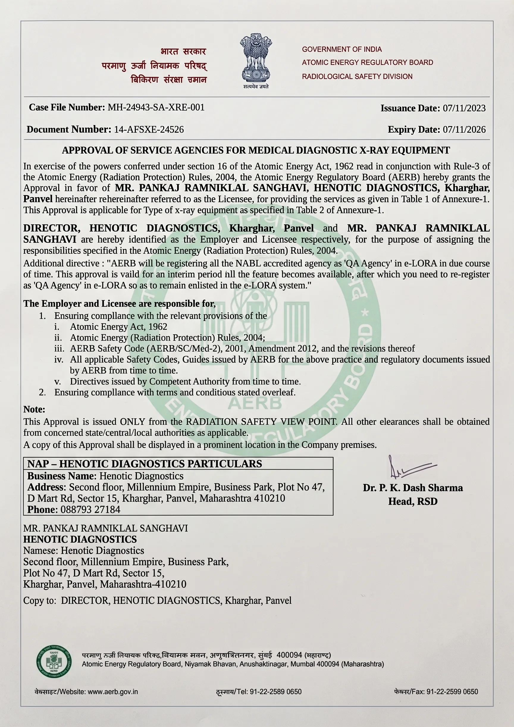
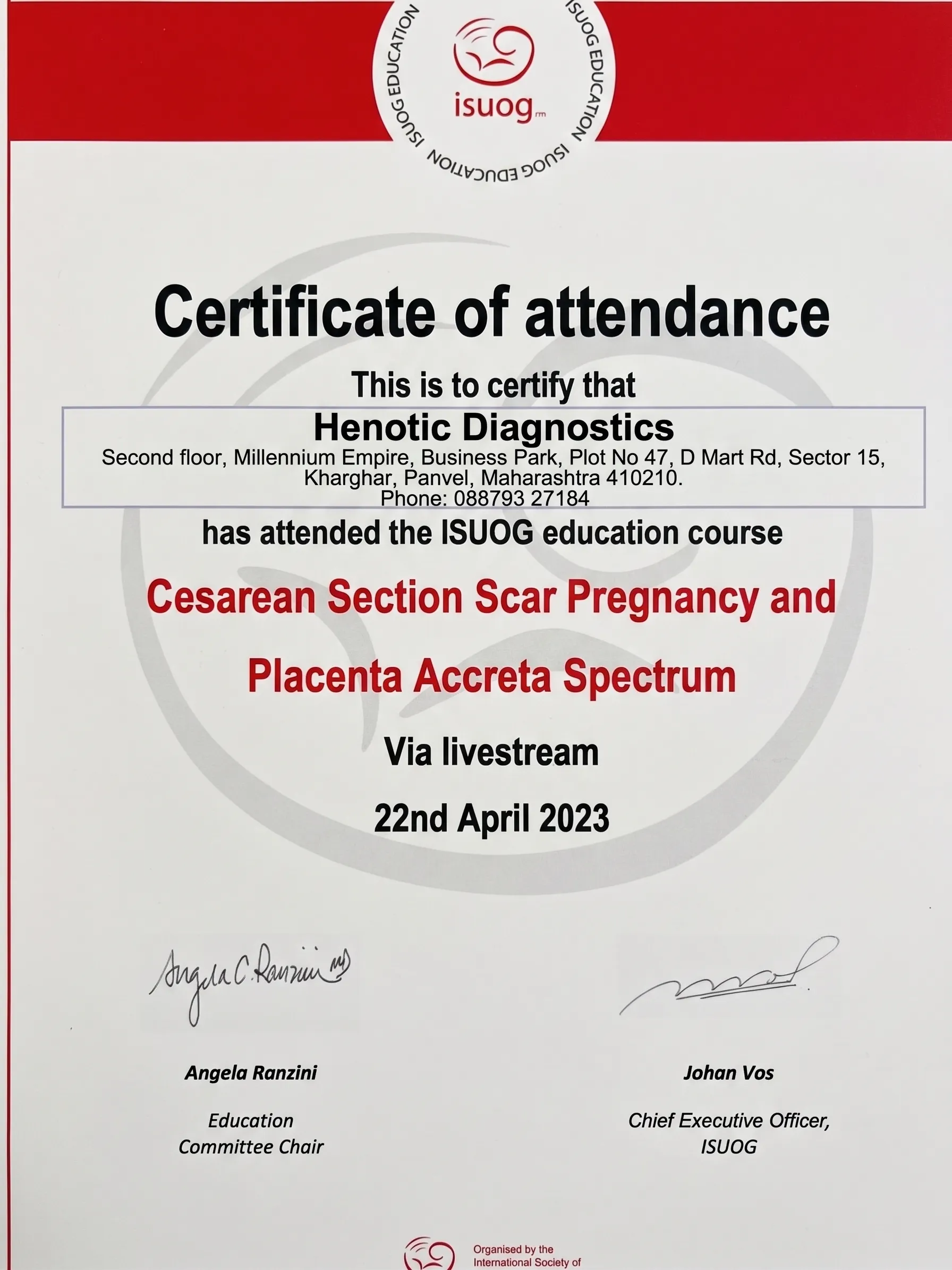
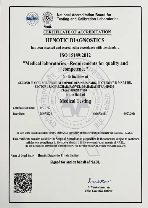
Specifically, Henotic Diagnostics Radiology Center maintains rigorous clinical quality control, radiation safety compliance, and government registration across all diagnostic imaging modalities.
.webp)





Furthermore, our official accreditation certificates demonstrate compliance with international ISO standards, atomic radiation safety guidelines, and clinical diagnostic benchmarks.
Specifically, read how advanced 3.0T MRI, 128-slice CT, and ultrasound diagnostic imaging at Henotic Diagnostics Radiology Center delivered accurate clinical clarity and compassionate care for our patients.
“Specifically, I was extremely anxious about my lumbar spine MRI scan due to severe claustrophobia. However, the 3.0T wide-bore MRI system at Henotic Diagnostics Radiology Center with quiet silent scan technology made the procedure completely comfortable. Furthermore, Dr. Pratibha Patil’s detailed reporting identified my L4-L5 disc herniation within hours. Consequently, my spine specialist could start treatment immediately!”
“In addition, my referring physician advised an urgent HRCT chest scan for persistent pulmonary symptoms. The 128-slice CT scan at Henotic Diagnostics was completed in less than 5 minutes with minimal radiation. Moreover, I received the verified digital report and DICOM images directly on WhatsApp on the same evening. As a result, the diagnostic clarity gave our family huge peace of mind!”
“Additionally, we visited Henotic Diagnostics Radiology Center for my Level 2 detailed fetal anomaly scan during the second trimester. The radiologist explained every anatomical structure on the screen with immense patience and care. Furthermore, the 4D ultrasound clarity was unbelievable. Therefore, we highly recommend their PCPNDT-registered sonography suite to all expecting parents!”
“Similarly, my father required a whole-body 18F-FDG PET-CT scan for oncology treatment monitoring. The reception staff and nuclear medicine technicians treated us with wonderful empathy and courtesy throughout intake. Consequently, the high-resolution PET-CT report provided accurate tumor margin evaluation for his oncologist. Truly an exceptional radiology center!”
Specifically, here are medically verified answers to common questions regarding diagnostic imaging modalities, radiation safety, scan preparation, and clinical indications.
Specifically, a radiology center is a specialized medical facility equipped with advanced imaging technologies—such as magnetic resonance imaging (MRI scan services), computed tomography (CT scan procedures), diagnostic ultrasound and sonography, PET-CT imaging, digital digital mammography, and X-ray—to non-invasively visualize internal body organs, soft tissues, bone structures, and blood flow. Consequently, radiology centers provide objective anatomical and physiological data used by physicians to diagnose diseases, evaluate traumatic injury, plan surgeries, and monitor medical treatments.
Additionally, a comprehensive radiology center provides a wide spectrum of diagnostic imaging modalities. Furthermore, these include 3.0T wide-bore MRI, 128-slice CT scans, whole-body PET-CT oncology scans, general and 4D obstetric ultrasound, Color Doppler study vascular evaluations, digital 3D mammography, DEXA bone mineral densitometry, FibroScan liver elastography, nuclear medicine scans, and image-guided biopsy/FNAC interventional procedures.
Neither modality is universally better; for instance, their clinical suitability depends on the anatomical organ and diagnostic question. Specifically, MRI scan procedures excel at evaluating soft tissues, brain parenchyma, spinal cord, nerve roots, joint cartilage, and pelvic organs without using ionizing radiation. In contrast, CT scan technology excels at rapid imaging of skeletal bone fractures, acute intracranial hemorrhage, chest/lung tissue, abdominal trauma, and complex 3D vascular angiography.
Indeed, yes, MRI is considered extremely safe because it utilizes powerful magnetic fields and radiofrequency pulses rather than ionizing radiation. However, because of the strong magnetic environment, detailed safety screening is performed prior to entering the scanner room to verify that patients do not have incompatible cardiac pacemakers, metallic implants, vascular clips, or metallic fragments.
Specifically, yes, Computed Tomography (CT) utilizes controlled ionizing X-rays to acquire cross-sectional slice images of internal body structures. Therefore, modern diagnostic radiology facilities utilize calibrated multi-slice scanners programmed with low-dose protocols adhering to ALARA (As Low As Reasonably Achievable) safety principles to minimize radiation while maintaining diagnostic clarity.
In fact, no, diagnostic ultrasound does not use ionizing radiation. Specifically, it operates using high-frequency, non-ionizing acoustic sound waves emitted by a specialized transducer and reflected back by tissue interfaces, making it completely safe for pregnancy monitoring, fetal development tracking, and routine abdominal evaluations.
Furthermore, a PET-CT scan procedure combines Positron Emission Tomography metabolic imaging with Computed Tomography anatomical mapping. Specifically, it is primarily used in oncology to detect primary malignant tumors, assess cancer staging, evaluate response to chemotherapy or radiotherapy, detect disease recurrence, and investigate specific cardiac or neurological disorders.
Additionally, a Color Doppler scan is a specialized ultrasound examination that evaluates blood circulation through arteries and blood vessels. Specifically, by calculating sound wave frequency shifts from moving red blood cells, Doppler imaging assesses arterial blockages, carotid artery stenosis, deep vein thrombosis (DVT), varicose veins, and fetal-placental blood flow in high-risk pregnancies.
For example, a 3T MRI is a 3.0 Tesla magnetic resonance imaging system operating at twice the magnetic field strength of standard 1.5T scanners. Consequently, this higher signal-to-noise ratio in our 3T MRI scan system delivers finer spatial resolution, shorter scan times, and enhanced sub-millimeter visualization of small anatomical structures in neuro, joint, and pelvic imaging.
Specifically, HRCT (High-Resolution Computed Tomography) is a specialized CT imaging technique using sub-millimeter slice thicknesses (typically 0.625mm to 1.25mm) and high-spatial-frequency reconstruction algorithms. As a result, it captures detailed lung parenchyma images during an HRCT Chest scan for evaluating interstitial lung disease (ILD), pulmonary fibrosis, bronchiectasis, and viral lung infections.
Moreover, mammography is a targeted low-dose X-ray examination of the breast designed to identify microcalcifications, small masses, and tissue distortions long before clinical symptoms occur. Specifically, performing 3D mammography (tomosynthesis) captures multiple low-dose images from different angles for cross-sectional breast evaluation.
In addition, DEXA (Dual-Energy X-ray Absorptiometry) is the gold-standard diagnostic imaging technique for measuring Bone Mineral Density (BMD). Specifically, it utilizes two low-dose X-ray beams to quantify bone loss, diagnose osteopenia and osteoporosis, and evaluate fracture risk at the lumbar spine and hip joints.
Similarly, FibroScan (Transient Elastography) is a non-invasive ultrasound-based examination that measures liver stiffness (in kilopascals) and quantifies hepatic fat content (CAP score). Therefore, it provides a quick, painless evaluation of liver fibrosis, cirrhosis, non-alcoholic fatty liver disease (NAFLD), and chronic hepatitis without needing a liver biopsy.
Finally, an image-guided biopsy is a minimally invasive diagnostic procedure where a radiologist uses real-time ultrasound or CT imaging to guide a thin needle precisely to a targeted tissue mass or lesion. Specifically, this extracts cellular samples (FNAC) or core tissue samples for histopathological laboratory analysis.
Specifically, if you require diagnostic imaging or radiology scan scheduling, do not delay. In contrast, contact our central diagnostic reception team to reserve your appointment.
Second floor, Millennium Empire, Business Park, Plot No 47, D Mart Rd, Sector 15, Kharghar, Navi Mumbai, Panvel, Maharashtra 410210
Specifically, providing tertiary hospital-grade medical imaging services across 3.0T MRI, 128-slice CT, PET-CT, ultrasound, color Doppler, digital mammography, nuclear medicine, DEXA, and FibroScan—backed by sub-specialty radiologist reporting and rapid digital PACS report delivery.
Get priority booking through our secure portal.