What Is a Medical Imaging Center?
What Is Medical Imaging?
In addition, medical imaging refers to the techniques and processes used to create visual representations of the interior of a body for clinical analysis and medical intervention. Consequently, at our Diagnostic Center, it encompasses non-invasive anatomical mapping, metabolic assessment, and functional tissue tracking.
How Does Medical Imaging Work?
For instance, imaging technologies utilize physical energy forms—including strong magnetic fields and radio waves (MRI Scan), high-frequency sound waves (Ultrasound), ionizing X-rays (CT Scan), and injected radiotracers (PET Scan)—to capture tissue densities and physiological signals.
What Can Medical Imaging Show?
As a result, scans conducted at a modern Medical Imaging Center reveal dense bone fractures, internal organ morphology, soft tissue injuries, vascular blood flow dynamics, fluid accumulations, cellular metabolic activity, and heart chamber motion.
Medical Imaging Center: Medical Imaging vs Diagnostic Testing
A Medical Imaging Center captures spatial structural and anatomical images of internal organs, whereas blood pathology laboratory diagnostic testing measures biochemical fluid markers and electrophysiological tests (ECG Test) measure electrical conduction.
Moreover, at Henotic Diagnostics, our Medical Imaging Center integrates hospital-grade imaging infrastructure with sub-specialty radiologist expertise. In particular, by capturing cross-sectional, 3D, and real-time functional data, our center equips referring physicians with actionable diagnostic clarity across oncology, neurology, cardiology, orthopedics, gastroenterology, and fetal medicine. According to the Radiological Society of North America (RadiologyInfo), proper imaging selection accelerates clinical diagnosis while minimizing unnecessary invasive procedures.
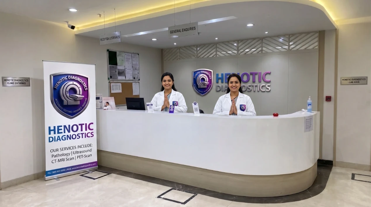
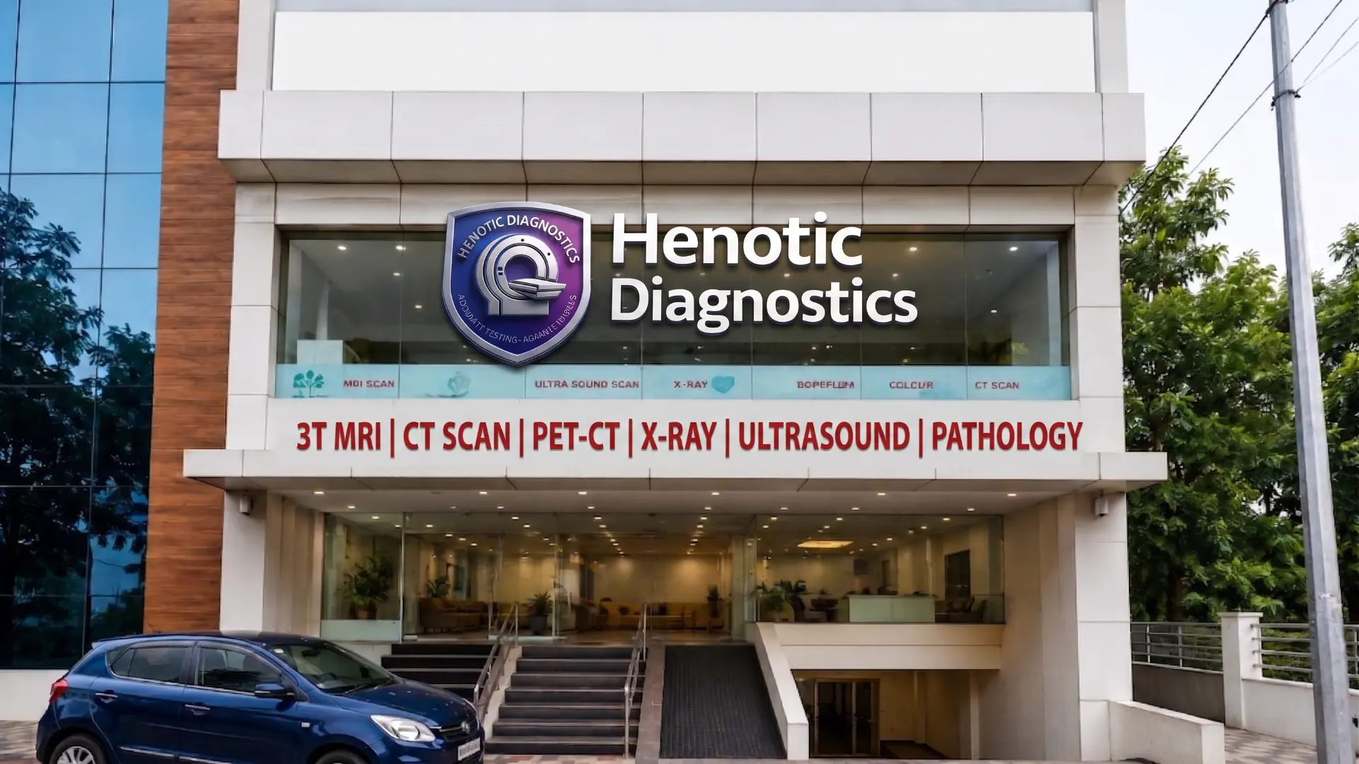
For example, Henotic Diagnostics Outpatient Medical Imaging Center Environment
Why Patients & Referring Clinicians Choose Henotic Diagnostics Medical Imaging Center
Additionally, delivering uncompromising radiological precision, advanced technology infrastructure, and compassionate patient care designed for diagnostic certainty.
Medical Imaging Center: Hospital-Grade Technology
Notably, our Medical Imaging Center operates tertiary hospital-grade imaging infrastructure featuring 3.0 Tesla wide-bore MRI, 128-slice sub-millimeter CT scanners, and Voluson E10 4D fetal ultrasound platforms calibrated daily for sub-millimeter anatomical resolution.
Medical Imaging Center: Sub-Specialty Radiologist Reporting
Therefore, every scan at our Medical Imaging Center is interpreted by board-certified radiologists with dedicated sub-specialty expertise in neuroradiology, musculoskeletal imaging, cardiac CT angiography, and fetal medicine, providing referring doctors with diagnostic clarity.
Medical Imaging Center: AERB & NABL Quality Accreditation
Indeed, our Medical Imaging Center strictly adheres to Atomic Energy Regulatory Board (AERB) radiation safety mandates, PCPNDT regulatory guidelines, and NABL laboratory accreditation standards, enforcing low ALARA radiation protocols.
Medical Imaging Center: Rapid Digital PACS Report Access
In fact, we expedite critical treatment decisions by offering rapid diagnostic report delivery. Overall, clinicians and patients gain immediate cloud access to high-resolution DICOM images and digital PDF reports through our secure online portal.
Medical Imaging Center: Ambient Patient-Centric Experience
Essentially, our Medical Imaging Center prioritizes patient comfort with wide-bore MRI systems, silent scan acoustic software, ambient lighting, and dedicated staff who assist anxious or pediatric patients through their imaging procedure.
Medical Imaging Center: Comprehensive Multi-Disciplinary Integration
Simultaneously, offering complete diagnostic services under one roof—integrating advanced radiology (MRI, CT, PET CT, USG Scan) with blood pathology, 2D Echo cardiology, FibroScan, and DEXA Scan—enabling thorough multi-system clinical correlation.
Types of Tests at Our Medical Imaging Center
Presently, Henotic Diagnostics Medical Imaging Center encompasses multiple specialized modalities, each operating on distinct physical principles to target specific tissue types and medical conditions:
Medical Imaging Center: MRI (Magnetic Resonance Imaging)
Likewise, uses magnetic fields and radiofrequency waves to generate non-ionizing images of soft tissues, brain micro-structures, spinal cord, and joint ligaments at our Medical Imaging Center.
Medical Imaging Center: CT Scan (Computed Tomography)
To clarify, combines rotating X-ray beams with multi-detector computing to reconstruct detailed cross-sectional 3D slices of internal organs, lung parenchyma, coronary arteries, and bone fractures.
Medical Imaging Center: Ultrasound & Sonography
Accordingly, employs high-frequency acoustic sound waves to visualize real-time dynamic motion within abdominal organs, pelvic structures, soft tissue nodules, and fetal development.
Medical Imaging Center: Doppler Ultrasound
Thus, specialized ultrasound evaluation of red blood cell motion, providing real-time color flow maps and velocity measurements across arteries, deep veins, and umbilical vessels.
Medical Imaging Center: X-Ray & Digital Radiography
With this in mind, projects quick, low-dose electromagnetic energy through body structures to produce immediate high-contrast diagnostic images of skeletal bones, chest fields, and joint alignment.
Medical Imaging Center: Mammography & Breast Imaging
Low-energy X-ray examination specifically calibrated for breast tissue, capturing 2D digital images and 3D tomosynthesis slices to detect microcalcifications and subtle lesions early.
Medical Imaging Center: PET-CT & Nuclear Medicine
Systematically, fuses functional cellular metabolic PET imaging (using 18F-FDG radiotracer) with structural CT anatomical localization for precise cancer staging and metabolic disease tracking.
Cardiac Imaging
Specifically, combines non-invasive 2D Echocardiography, CT Coronary Angiography, TMT stress testing, and Cardiac MRI to analyze myocardial wall motion, valvular function, and electrical conduction.
DEXA / Bone Density Scan
Furthermore, dual-Energy X-Ray Absorptiometry non-invasively quantifies bone mineral density (BMD) at the lumbar spine and hip to diagnose osteoporosis and assess fracture risk.
FibroScan / Liver Elastography
In addition, transient elastography non-invasively measures shear wave propagation velocity through hepatic tissue to quantify liver stiffness and fat content without liver biopsy.
Digital Imaging, PACS & Medical Imaging Center Report Access
What Is PACS?
Consequently, picture Archiving and Communication System (PACS) is a medical imaging technology used at our Medical Imaging Center to securely store, retrieve, manage, and share electronic DICOM images.
Digital DICOM Imaging
For instance, high-resolution DICOM standard imaging files capture sub-millimeter volume data, enabling multi-planar 3D reconstruction and precise radiologist measurement.
Online Imaging Reports
As a result, patients receive secure digital portal access to official radiologist PDF diagnostic reports and high-resolution imaging studies following verification.
Sharing Studies With Doctors
Moreover, encrypted digital cloud links allow seamless sharing of DICOM imaging studies directly with your attending physician or surgeon for rapid consultation.
Medical Imaging Services at Henotic Diagnostics Medical Imaging Center
Magnetic Resonance Imaging (3T MRI Services)
In particular, magnetic Resonance Imaging (MRI) employs powerful magnetic fields and high-frequency radio waves to generate high-contrast multi-planar images of soft tissues, nerves, muscles, ligaments, and deep organ structures without utilizing ionizing radiation. For example, Henotic Diagnostics Medical Imaging Center operates a state-of-the-art 3 Tesla (3T) MRI scanner offering superior signal-to-noise ratio, ultra-high spatial resolution, and accelerated scan times.
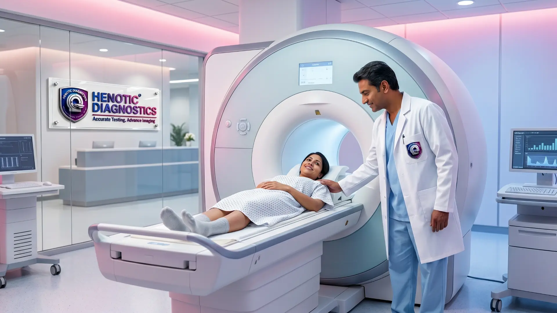
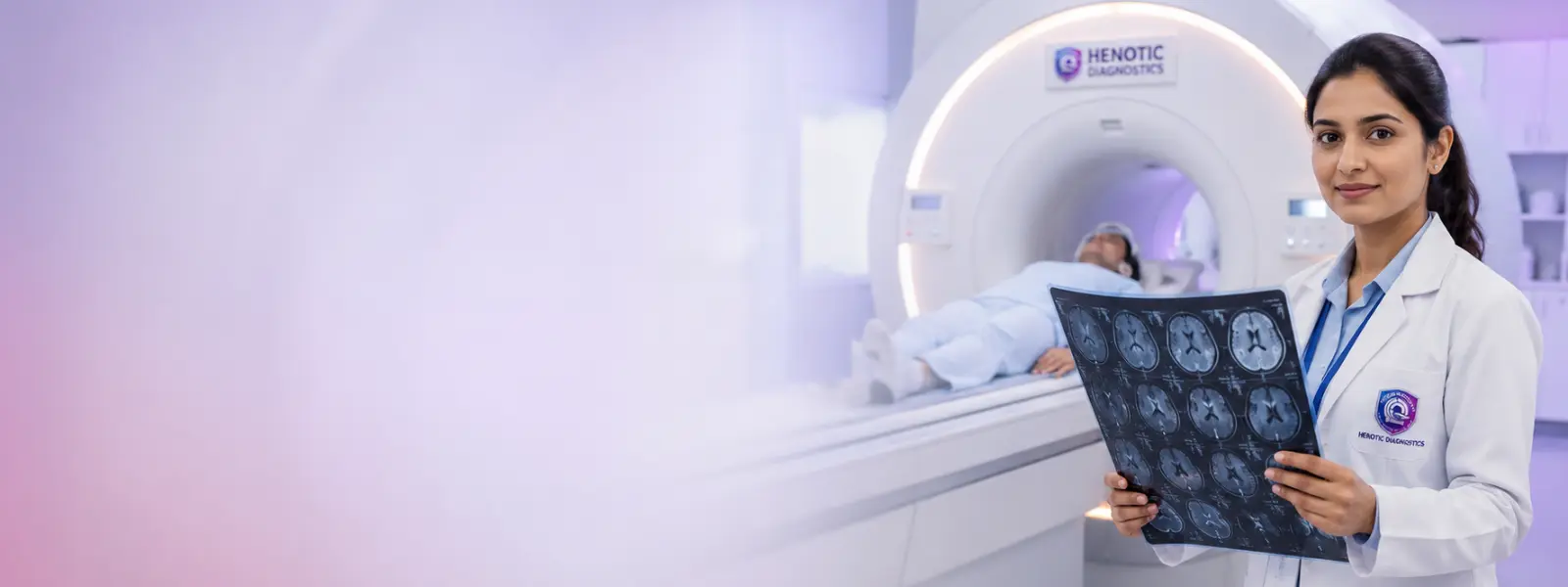
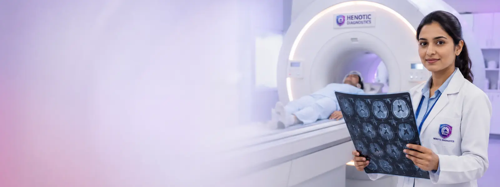
Additionally, 3.0T High-Field Magnetic Resonance Imaging System at Medical Imaging Center
| Ionizing Radiation | Contrast Agent | Typical Duration | Primary Target Systems |
| NONE (0 mSv) | Gadolinium (Optional) | 20 – 45 Minutes | Brain, Spine, Joints, Pelvis, Biliary System |
Notably, explore dedicated modality page: 3T MRI Scan Services
Computed Tomography (128-Slice CT Scan Services)
Therefore, computed Tomography (CT) combines specialized rotating X-ray equipment with powerful computer algorithms to construct detailed cross-sectional “slices” of internal vascular structures, abdominal organs, chest parenchyma, and skeletal bone morphology. Indeed, our 128-slice CT scanner delivers rapid sub-millimeter volume coverage, facilitating high-speed cardiac coronary angiography and low-dose screening protocols.
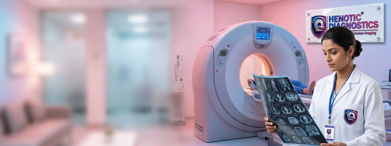
In fact, advanced 128-Slice Computed Tomography Suite at Medical Imaging Center
| Ionizing Radiation | Contrast Agent | Typical Duration | Primary Target Systems |
| Low-Dose X-Ray (ALARA) | Iodinated Media (Optional) | 5 – 15 Minutes | Lungs (HRCT), Coronary Arteries, Bone Fractures, Trauma |
Overall, explore dedicated modality page: 128-Slice CT Scan Services
Diagnostic Ultrasound & Sonography Services
Essentially, diagnostic Ultrasonography (USG) uses non-ionizing high-frequency sound waves to capture real-time dynamic images of soft tissues, fluid accumulations, internal organ structures, and blood flow velocity. Simultaneously, ultrasound is radiation-free, making it the ideal diagnostic modality for abdominal organ evaluation, pelvic screening, thyroid nodule characterization, and pediatric diagnostics.

Presently, radiation-Free Diagnostic Ultrasonography Suite at Medical Imaging Center
Likewise, verified Services: Abdominal Ultrasound, Pelvic Ultrasound, Kidney USG, Thyroid Ultrasound, Breast Ultrasound, Prostate Ultrasound, Upper Abdomen Sonography, and Ultrasound-Guided Procedure.
To clarify, explore dedicated modality page: Ultrasound & Sonography Services
Color Doppler Vascular Imaging Services
Accordingly, color Doppler ultrasound evaluates vascular hemodynamics, converting high-frequency sound wave frequency shifts into vivid color-coded maps of red blood cell flow velocity and direction. Thus, doppler imaging is critical for diagnosing deep vein thrombosis (DVT), arterial stenoses, carotid artery plaques, varicose veins, and organ graft perfusion.
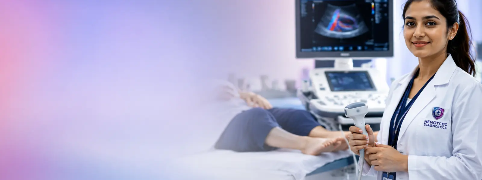
With this in mind, vascular Hemodynamic Color Doppler System at Medical Imaging Center
Systematically, verified Services: Arterial Doppler, Venous Doppler, Carotid Color Doppler, Renal Artery Doppler, DVT Doppler, and Obstetric Pregnancy Doppler.
Specifically, explore dedicated modality page: Color Doppler Scan Services
Advanced Digital Radiography & X-Ray Services
Furthermore, modern X-Ray imaging projects low-dose electromagnetic energy through body structures to produce immediate high-contrast diagnostic images of dense bone structures, joint spaces, and lung fields. Furthermore, state-of-the-art digital detector arrays allow lower radiation exposure, enhanced contrast adjustments, and immediate digital archiving.
In addition, low-Dose Digital Radiography Suite at Medical Imaging Center
Consequently, verified Services: Digital Chest X-Ray (PA/Lateral), Spine Radiography (Cervical/Lumbar), Bone Fracture X-Rays, Joint Radiography, and Abdominal Flat-Plate Radiographs.
For instance, status: PLANNED PAGE — URL NOT VERIFIED
Mammography & Advanced Breast Imaging
Digital Mammography utilizes low-energy X-ray beams optimized specifically to inspect breast tissue architecture for microcalcifications, subtle masses, asymmetric densities, and early non-palpable malignant lesions. As a result, our center provides Digital Mammography, Digital Breast Tomosynthesis (DBT), and complementary Sonomammography. Moreover, guidelines from the National Cancer Institute (NCI) emphasize early mammographic screening for optimal prognosis.
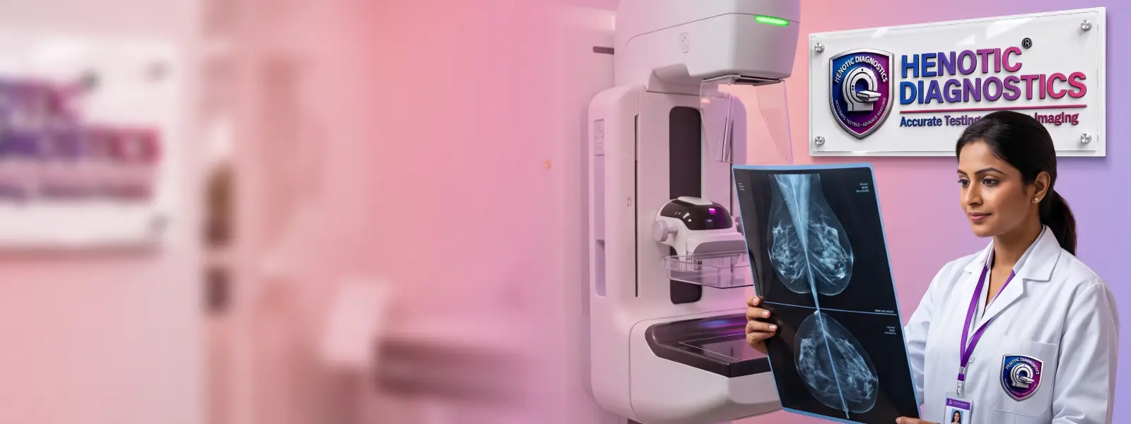
In particular, digital Breast Tomosynthesis & Mammography Suite at Medical Imaging Center
For example, explore dedicated modality page: Digital Mammography Services
Obstetric, Pregnancy & Fetal Imaging Services
Additionally, fetal diagnostic imaging provides radiation-free monitoring of embryonic development, anatomical structural integrity, placental positioning, amniotic fluid volume, and umbilical blood flow. Notably, performed by certified sonologists under strict PCPNDT regulatory compliance, our fetal imaging suite provides comprehensive maternal-fetal surveillance.
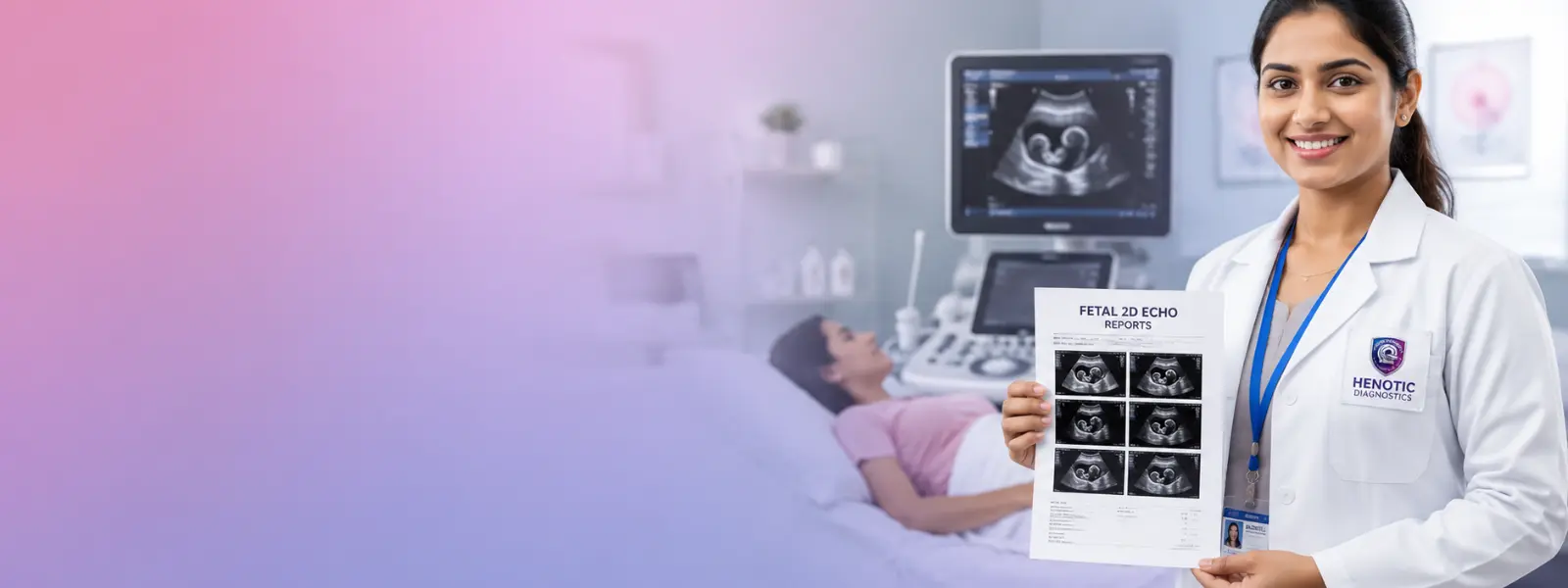
Therefore, pCPNDT-Compliant Fetal & Obstetric Suite at Medical Imaging Center
Verified Services: Early Pregnancy Viability Scan, First Trimester NT Scan, Level II Anomaly Scan, Fetal Echo, Fetal Growth Scan, Follicular Study, and HSG Test.
PET-CT & Molecular Nuclear Medicine Services
Indeed, positron Emission Tomography–Computed Tomography (PET-CT) fuses functional metabolic imaging (PET) with precise structural anatomical localization (CT). By administering radiotracer analogues such as Fluorodeoxyglucose (18F-FDG PET-CT), PET-CT detects hyper-metabolic neoplastic activity, assesses cancer staging, monitors treatment response, and localizes occult inflammatory processes.

In fact, molecular PET-CT Cancer Imaging Platform at Medical Imaging Center
Overall, verified Services: DOTA PET Scan, DOPA PET CT, PSMA PET Scan, Bone Scans, Thyroid Scans, Renal Scans, DTPA Scans, and Myocardial Perfusion Scans.
Essentially, explore dedicated modality pages: PET Scan Services | PET-CT Services
Non-Invasive Cardiac Imaging & Functional Diagnostics
Simultaneously, cardiovascular imaging combines dynamic ultrasound, electrophysiology, and exercise stress testing to non-invasively analyze cardiac chamber dimensions, valvular competence, myocardial wall motion, and electrical conduction. Presently, according to the World Health Organization (WHO), early non-invasive cardiac evaluation is essential for preventing major cardiovascular events.
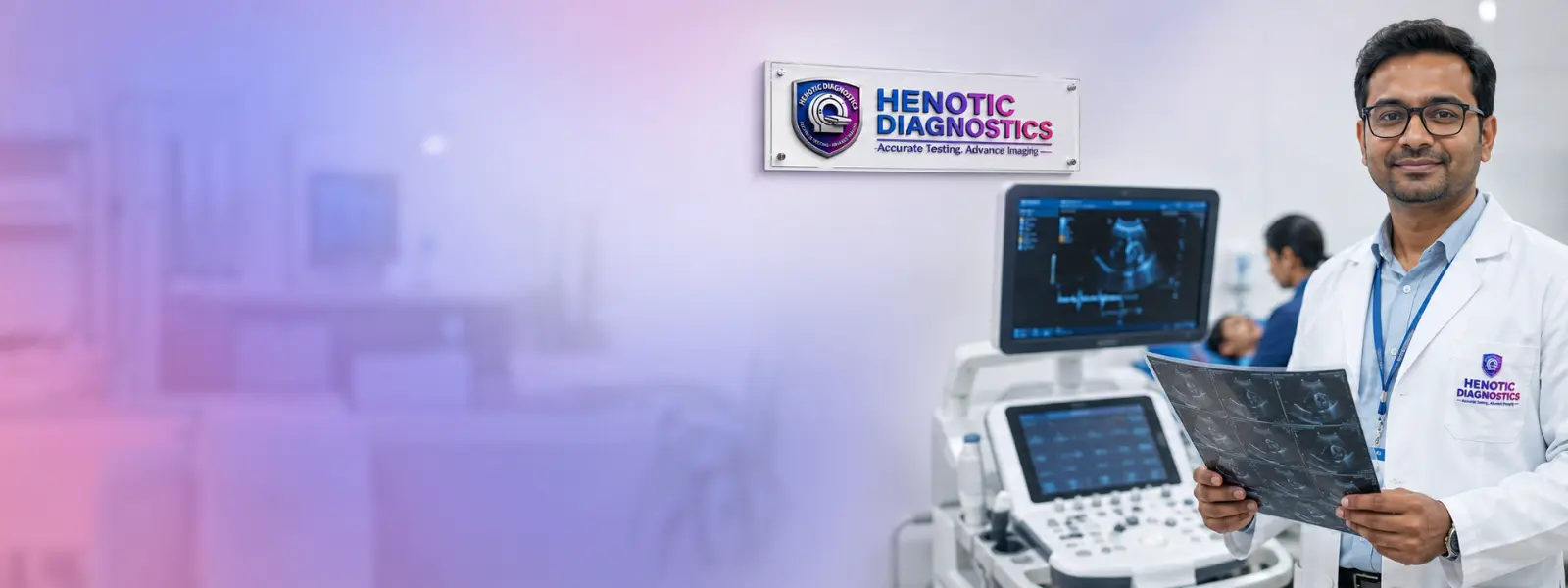
Likewise, non-Invasive Cardiovascular Diagnostics Suite at Medical Imaging Center
To clarify, verified Services: 2D Echocardiography, ECG Testing, TMT Treadmill Stress Test, and Holter Monitoring.
Specialized Liver & Bone Density Imaging (FibroScan & DEXA)
Accordingly, advanced specialized diagnostic equipment provides quantitative tissue characterization. Thus, fibroScan (Liver Elastography) non-invasively measures liver shear wave speed to quantify hepatic fibrosis and steatosis without liver biopsy. With this in mind, DEXA (Dual-Energy X-Ray Absorptiometry) accurately quantifies bone mineral density (BMD) to diagnose osteoporosis and estimate fracture risk.
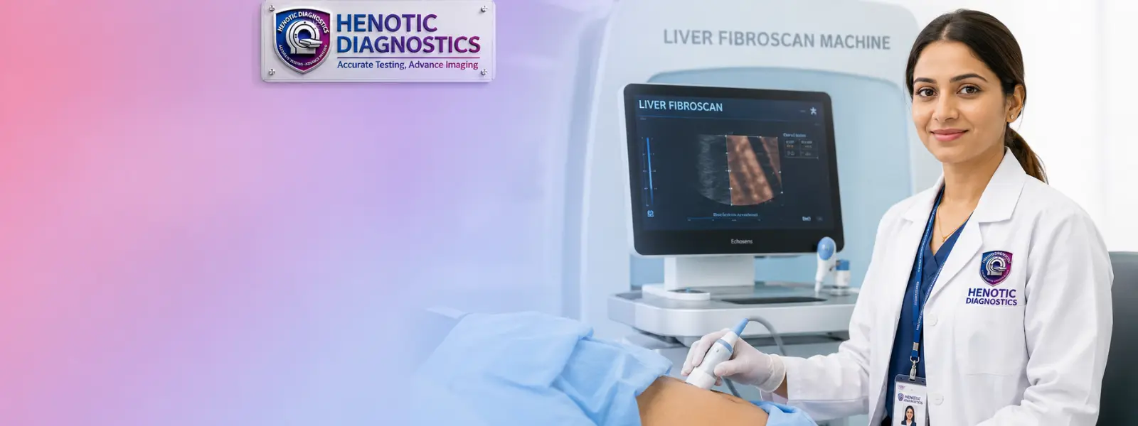
Systematically, quantitative FibroScan & DEXA Bone Mineral Density Suite at Medical Imaging Center
Advanced Technologies at Our Medical Imaging Center
3 Tesla MRI
Specifically, ultra-high magnetic field strength providing sub-millimeter spatial resolution and high signal-to-noise ratio for brain micro-structure and joint imaging at our Medical Imaging Center.
128-Slice CT
Multi-detector volume coverage capturing 128 slices per gantry rotation, enabling sub-second coronary angiography and low-dose lung screening.
Digital Radiography
Furthermore, flat-panel digital detectors replacing film arrays to reduce radiation exposure and deliver instantaneous high-contrast bone and chest images.
3D Mammography / Digital Breast Tomosynthesis
In addition, multi-angle low-dose X-ray sweeps creating 1mm 3D breast tissue slice reconstructions to uncover hidden microcalcifications in dense breasts.
Advanced Ultrasound & Color Doppler
Consequently, Voluson E10 4D probes and high-frequency Doppler arrays for real-time fetal biometry and hemodynamic vascular flow mapping.
PET-CT Imaging
For instance, molecular nuclear medicine suite combining 18F-FDG metabolic radiotracer uptake with anatomical CT mapping for early cancer staging.
Digital Image Archiving & PACS
As a result, high-security Picture Archiving and Communication Systems enabling instant digital cloud archiving and DICOM access for referring physicians.
Medical Imaging Center Services by Clinical Specialty
Neurology & Brain Imaging
Moreover, 3T Brain MRI, CT Brain, Cerebral Angiography, and Carotid Doppler for stroke, epilepsy, tumors, and neurodegenerative disorders.
Orthopedic & Musculoskeletal Imaging
In particular, 3T MRI Joint Scans, Whole Spine MRI, Digital X-Rays, and DEXA Scan for fractures, ligament tears, and arthritis.
Cardiac & Cardiovascular Imaging
For example, CT Coronary Angiography, 2D Echocardiography, TMT Stress Test, and Holter Monitoring for coronary disease.
Pulmonary & Chest Imaging
Additionally, HRCT Chest, Low-Dose CT Lung Screening, Chest X-Rays, and PET-CT for lung nodules, pneumonia, and ILD.
Gastrointestinal & Abdominal Imaging
Notably, triple-phase CT Abdomen, Abdominal 3T MRI, MRCP Scan, and Abdominal Ultrasound for pancreatitis and gallstones.
Women’s Health & Breast Imaging
Therefore, digital Mammography, 3D Mammography, Sonomammography, and Pelvic USG for gynecological wellness.
Pregnancy & Fetal Imaging
Indeed, early Pregnancy Scan, NT Scan, Anomaly Scan, Fetal Echo, and Growth Scan surveillance.
Oncology & Cancer Imaging
In fact, whole Body FDG PET-CT, Nuclear Bone Scan, Contrast MRI, and CT staging for early cancer detection.
Liver & Hepatobiliary Imaging
Overall, fibroScan Liver Elastography, MRCP biliary mapping, Abdominal USG, and multiphase liver CT for fatty liver.
Urological & Kidney Imaging
Essentially, non-contrast CT KUB, Kidney USG, Renal Doppler, Prostate Ultrasound, and Pelvic MRI for urinary tract disease.
Medical Imaging Center Diagnostic Scans by Anatomical Body Part
Brain & Neurological System
Simultaneously, 3T Brain MRI, Stroke Protocol MRI, CT Brain, CT Cerebral Angiography, Carotid Color Doppler.
Spine & Musculoskeletal System
Presently, whole Spine 3T MRI, Joint Ligament MRI, Digital Bone X-Rays, DEXA Bone Density Screening.
Heart & Cardiovascular System
Likewise, CT Coronary Angiography, 2D Echo, Cardiac MRI, TMT Stress Test, Holter Monitoring.
Chest & Lungs
To clarify, HRCT Chest, Low-Dose CT Lung Screening, Chest Digital X-Ray, FDG PET-CT Lung Evaluation.
Liver & Gallbladder
Accordingly, fibroScan Liver Elastography, MRCP Biliary Mapping, Abdominal Ultrasound, Multiphase CT.
Kidneys & Urinary System
Thus, CT KUB, Kidney Ultrasound, Renal Doppler, Prostate Ultrasound, Pelvic MRI.
Thyroid & Neck
With this in mind, thyroid Ultrasound, Carotid Color Doppler, Soft Tissue Neck CT/MRI.
Breast
Systematically, digital Mammography, Digital Breast Tomosynthesis, Breast Ultrasound, Breast MRI.
Pelvis & Reproductive Organs
Specifically, pelvic 3T MRI, Transvaginal Ultrasound (TVS), Follicular monitoring, Prostate USG.
Vascular & Peripheral Circulation
Furthermore, arterial Doppler, Venous Doppler, DVT Doppler, CT Angiography.
In addition, clinical Distinction: Screening imaging is intended for people without known disease or symptoms in appropriate populations; diagnostic imaging is performed to investigate specific symptoms, clinical signs, or known medical conditions.
Screening Imaging
Consequently, evaluates healthy, asymptomatic individuals to uncover sub-clinical pathology early (e.g., annual screening mammography, low-dose CT lung screening in high-risk groups).
Diagnostic Imaging
For instance, investigates active symptoms (pain, neurological deficits, abnormal lab values) to pinpoint anatomical pathology and direct medical treatment at our Medical Imaging Center.
Imaging for Treatment Monitoring
Evaluates therapeutic efficacy during or following clinical treatment (such as measuring tumor metabolic response on serial FDG PET-CT scans).
Imaging for Follow-Up
As a result, provides long-term disease surveillance post-recovery to confirm structural stability and rule out disease recurrence over time.
When Is Medical Imaging Recommended?
Imaging for Symptoms
Recommended when patients present with persistent symptoms such as severe headache, abdominal pain, joint swelling, or chronic cough.
Imaging for Screening
Moreover, recommended for age-appropriate health checkups, high-risk cancer screening, or routine wellness surveillance.
Imaging for Injury and Trauma
In particular, urgent evaluation following physical trauma, sports injuries, or motor accidents to rule out internal hemorrhage or bone fractures.
Imaging for Sudden or Emergency Symptoms
Rapid diagnostic assessment during acute medical emergencies such as suspected acute stroke, pulmonary embolism, or acute abdomen.
Imaging for Follow-Up
For example, scheduled periodic scans to track known chronic medical conditions, post-operative healing, or therapeutic responses.
Imaging Before or After Treatment
Additionally, pre-surgical anatomical mapping or post-chemotherapy baseline evaluations to guide surgical and medical management.
How to Choose the Right Medical Imaging Test
Key Clinical Decision Criteria for Diagnostic Test Selection
Notably, selecting the appropriate diagnostic imaging modality involves balancing anatomical tissue composition, clinical urgency, radiation exposure considerations, and contrast media requirements:
- Soft Tissue vs. Bone Structures: MRI is superior for brain micro-structures, spinal cord, ligaments, and deep soft tissue, while CT is optimal for acute bone fractures, lung parenchyma, and rapid trauma evaluation.
- Radiation Considerations: Ultrasound and MRI involve no ionizing radiation, making them the preferred first-line imaging choice for pregnant patients, pediatric evaluations, and serial follow-up studies.
- Metabolic Activity vs. Anatomical Mapping: PET-CT evaluates cellular glucose metabolism for early oncological detection, whereas CT and MRI focus primarily on structural anatomy.
- Vascular Flow Dynamics: Color Doppler provides real-time dynamic blood flow velocity mapping without invasive arterial puncture.
MRI vs CT vs Ultrasound vs X-Ray
Medical Imaging Center: MRI vs CT Scan
Therefore, MRI uses magnets with zero radiation and excels in soft tissue/neuro contrast; CT uses X-rays, takes minutes, and excels in lung, bone fracture, and emergency trauma evaluation.
Medical Imaging Center: MRI vs Ultrasound
Indeed, both avoid ionizing radiation. In fact, ultrasound is portable and real-time for fluid/pelvis/fetal imaging; MRI provides deep 3D multi-planar tissue resolution across the whole body.
Medical Imaging Center: CT Scan vs X-Ray
Overall, x-Rays provide instant 2D projection images for routine chest/bone checks; CT combines hundreds of rotating X-rays into 3D cross-sectional volume slices.
Medical Imaging Center: Ultrasound vs CT Scan
Essentially, ultrasound uses sound waves without radiation, perfect for superficial soft tissues; CT penetrates deep bowel and retroperitoneal spaces using low-dose X-ray beams.
| Test | Uses | Ionizing Radiation | Common Strengths |
|---|---|---|---|
| MRI | Magnetic fields / Radiofrequency | No | Soft tissues, brain micro-structures, spine, joints |
| CT | X-rays | Yes | Rapid cross-sectional imaging, lungs, trauma, bone fractures |
| Ultrasound | Sound waves | No | Real-time soft tissue, abdominal/pelvic and obstetric imaging |
| X-ray | X-rays | Yes | Bones and chest screening |
Which Imaging Test Is Appropriate?
Simultaneously, your attending doctor determines the appropriate modality based on suspected pathology, anatomical site, radiation limits, and clinical urgency.
Contrast Media in Medical Imaging
What Is Contrast Media?
Presently, specialized chemical substances administered orally or intravenously to temporarily change how imaging signals interact with internal tissues.
Why Is Contrast Used?
Likewise, enhances tissue contrast, highlighting blood vessel boundaries, organ perfusion, inflammation, and tumor vascularity for diagnostic clarity.
MRI Contrast
To clarify, uses gadolinium-based contrast agents (GBCAs) which alter magnetic relaxation times to outline central nervous system lesions and vascularity.
CT Contrast
Accordingly, uses non-ionic iodinated contrast media that block X-ray beams to brightly map arterial blood flow, liver lesions, and urinary excretion.
Important Information Before Contrast Imaging
Depending on the examination, medical history, and local protocol, kidney-function assessment (such as Serum Creatinine testing) may be requested before contrast administration to assess kidney function and manage contrast-related risk.
Contrast Media Safety and Possible Reactions
Thus, modern contrast agents are exceptionally safe and rigorously tested. Most minor reactions—such as a brief flushing sensation, metallic taste, or mild nausea—resolve quickly on their own. With this in mind, severe allergic responses are exceedingly rare, and our Medical Imaging Center is fully equipped with advanced emergency monitoring and medical protocols to ensure patient safety.
Radiation Safety in Medical Imaging
What Does ALARA Mean?
Systematically, ALARA stands for “As Low As Reasonably Achievable,” a fundamental safety principle guiding radiation optimization at our Medical Imaging Center.
Which Imaging Tests Use Radiation?
Specifically, CT Scans, Digital X-Rays, Mammography, and PET-CT utilize low-dose ionizing radiation. Furthermore, MRI and Ultrasound involve zero radiation.
How Is Exposure Minimized?
In addition, calibrated detector arrays, beam collimation, anatomical shielding, and automated exposure control minimize radiation dose.
Are Repeated CT Scans Safe?
Consequently, repeated CT scans are not automatically risk-free; each repeat examination must be clinically justified by your attending doctor.
Radiation Safety for Children
For instance, pediatric protocols use weight-adjusted dose reductions to protect growing tissues during necessary diagnostic studies.
Medical Imaging During Pregnancy
Ultrasound During Pregnancy
As a result, ultrasound does not use ionizing radiation and is widely used during pregnancy when clinically appropriate for embryonic viability and fetal growth monitoring.
Doppler During Pregnancy
Moreover, obstetric Pregnancy Doppler uses sound waves to non-invasively evaluate placental blood flow velocity in uterine and umbilical arteries.
MRI During Pregnancy
In particular, MRI may be performed during pregnancy when clinically indicated; decisions depend on the specific clinical situation and imaging requirements.
When X-Ray or CT May Be Considered
For example, X-ray or CT examinations may be performed during pregnancy when medically justified for urgent maternal evaluation, utilizing appropriate abdominal shielding precautions.
Why You Should Tell the Imaging Team About Pregnancy
Informing your technologist and doctor beforehand allows the medical team to prioritize non-ionizing modalities (such as Ultrasound or MRI), apply specialized abdominal shielding, or optimize dose settings to protect developing tissues.
Additionally, discussing Imaging With Your Doctor: Always inform your technologist and physician prior to any diagnostic scan if you are pregnant or suspect you may be pregnant.
What Happens During a Medical Imaging Test?
Before the Scan
Notably, registration, metal screening, changing into hospital gowns, and establishing IV access if contrast media is indicated.
During the Scan
Comfortable positioning on the motorized scanner table, remaining still, and following automated breath-hold instructions.
Contrast Administration
Therefore, automated IV injection of gadolinium or iodinated contrast media mid-procedure, causing a brief warm sensation.
After the Scan
Indeed, immediate removal of IV lines, brief observation, resuming normal diet, and maintaining hydration to flush contrast.
How Long Does Medical Imaging Take?
In fact, x-rays take minutes; CT scans take 5–15 minutes; MRI scans take 20–45 minutes; PET-CT scans take 2–3 hours total including uptake time.
Is Medical Imaging Painful?
Overall, diagnostic imaging is non-invasive and painless. Minor temporary discomfort may arise from lying still or IV needle placement for contrast.
When Can I Return to Normal Activities?
Essentially, patients can immediately return to normal daily work, driving, and diet activities following routine diagnostic imaging scans.
How to Prepare for Different Medical Imaging Tests
Simultaneously, preparation requirements vary by examination. Presently, follow the specific instructions provided for your appointment at our Medical Imaging Center.
Medical Imaging Center: MRI Preparation
Likewise, remove all metallic items/jewelry. To clarify, fast 4 hours before contrast scans and bring recent Serum Creatinine lab reports if requested.
Medical Imaging Center: CT Scan Preparation
Accordingly, fast 4 to 6 hours before CECT contrast scans. Thus, wear comfortable clothing without metal zippers or buttons.
Medical Imaging Center: Ultrasound Preparation
With this in mind, abdominal USG requires 6-8 hours fasting. Systematically, pelvic/KUB USG requires drinking 1 liter of water 1 hour prior for a full bladder.
Medical Imaging Center: Doppler Preparation
Specifically, renal Doppler requires 6 hours fasting. Furthermore, limb vascular Doppler studies require no special dietary restrictions.
Medical Imaging Center: X-Ray Preparation
In addition, routine X-rays require zero dietary fasting. Consequently, remove metallic necklaces, belts, and pins from the target body region.
Mammography Preparation
For instance, do not apply deodorants, antiperspirants, talcum powders, or lotions under arms or breasts on the day of the exam.
PET-CT Preparation
As a result, strict 6-hour fasting before 18F-FDG injection. Moreover, maintain a low-carbohydrate diet and avoid strenuous exercise 24 hours prior.
Cardiac Imaging Preparation
In particular, for TMT stress tests, wear comfortable walking shoes. For example, avoid caffeine 12 hours before cardiac CT coronary angiography.
Medical Imaging Center Safety & Patient Considerations
MRI Safety and Metal Implants
Additionally, ferromagnetic metallic implants or foreign bodies must be pre-screened to prevent magnetic displacement.
Contrast Media Considerations
Notably, adverse contrast reactions are rare. Therefore, patients with previous drug allergies or asthma are pre-screened by medical staff.
Kidney Function and Contrast
Indeed, assessing kidney function via lab testing helps evaluate contrast clearance capability prior to administration.
Hearing Aids and Electronic Devices
In fact, hearing aids, glucose monitors, and external electronic devices must be removed prior to entering high magnetic field environments.
Pacemakers and MRI
Overall, traditional pacemakers preclude MRI. Essentially, modern MR-conditional pacemakers require specific cardiology programming protocols.
Tattoos and MRI
Simultaneously, dark tattoo inks containing metallic iron pigments may rarely experience slight warmth; cold compresses are applied if needed.
Claustrophobia During MRI
Presently, wide-bore 3T MRI systems, ambient lighting, and music headsets significantly reduce claustrophobic anxiety.
Allergies and Contrast History
Always report previous reactions to iodine or gadolinium contrast so appropriate pre-medication or alternative modalities can be selected.
Who Interprets Scans at a Medical Imaging Center?
Role of a Radiologist
Likewise, a radiologist is a specialized medical doctor (MD) trained in evaluating diagnostic imaging studies, identifying pathology, and authoring formal reports.
Subspecialty Radiology
Radiologists with subspecialty training focus on specific organ systems such as neuroradiology, musculoskeletal, cardiac, or fetal imaging.
How Radiologists Interpret Images
To clarify, radiologists analyze multi-planar cross-sectional DICOM image sequences, comparing tissue signal intensities and anatomical symmetry.
Radiology Report vs Clinical Diagnosis
Accordingly, the radiology report details objective imaging observations; your attending physician integrates these findings with your clinical exam to make a diagnosis.
Thus, why Clinical Correlation Matters: Imaging findings must always be correlated clinically with physical examination and lab results by your attending physician.
Understanding Medical Imaging Center Reports
With this in mind, a medical imaging report is an official clinical document generated by a qualified radiologist at our Medical Imaging Center following image analysis. Systematically, key sections include:
What Are “Findings”?
Specifically, detailed objective anatomical observations, measurements, and slice-by-slice organ descriptions recorded during radiologist interpretation.
What Is the “Impression”?
Furthermore, the radiologist’s synthesized diagnostic conclusion summarizing key findings into a definitive diagnostic statement for your doctor.
What Does “Clinical Correlation” Mean?
In addition, indicates that imaging observations must be evaluated alongside physical exams, patient history, and lab test results.
Who Should Explain My Imaging Report?
Your attending physician or specialist is best qualified to explain your imaging report in the context of your overall medical care.
Can an Imaging Report Diagnose a Disease by Itself?
Consequently, rarely. Diagnostic imaging provides essential anatomical data, but complete clinical diagnosis usually requires correlation with symptoms and lab tests.
Image-Guided Diagnostic Procedures
Ultrasound-Guided Procedures
For instance, real-time acoustic guidance for fluid aspirations, cyst drainage, and soft tissue needle placement tracking.
Image-Guided Tissue Sampling
As a result, minimally invasive biopsy sampling of thyroid nodules, breast masses, or liver lesions guided by real-time ultrasound or CT.
What Is an Image-Guided Biopsy?
Moreover, a procedure using live imaging to direct a biopsy needle into suspicious tissue to harvest cells for pathological analysis.
CT-Guided Procedures
In particular, precision cross-sectional CT navigation for deep thoracic or retroperitoneal tissue sampling.
When Image Guidance Is Used: Utilized whenever pinpoint accuracy is required to target internal lesions safely while completely avoiding adjacent blood vessels or organs.
Medical Imaging vs Medical Treatment
For example, medical imaging is primarily diagnostic—used to non-invasively visualize, characterize, detect, evaluate, or monitor internal pathological conditions. It is distinct from therapeutic medical treatments (such as surgical interventions, medication administration, or radiation therapy) that directly treat disease. Additionally, diagnostic imaging provides the essential visual roadmap that enables physicians to design precise treatment plans.
Senior Reporting Radiologist & Medical Leadership
Specifically, expert diagnostic imaging interpretation at Henotic Diagnostics Radiology Center is led by our distinguished board-certified consultant radiologist.

Dr. Pratibha Patil
Notably, MBBS, MD (Radiodiagnosis)
Clinical Leadership & Diagnostic Expertise
Dr. Therefore, Pratibha Patil serves as Lead Senior Reporting Radiologist at Henotic Diagnostics Radiology Center, dedicated to sub-specialty cross-sectional diagnostic imaging, neuro & musculoskeletal MRI interpretation, 128-slice CT angiography, and interventional sonography. Furthermore, she oversees our specialized diagnostic protocol optimization, ensuring high spatial resolution image acquisition for 3.0T wide-bore MRI, PET-CT molecular oncology mapping, and 4D obstetric ultrasound. Consequently, her detailed sub-specialty diagnostic reports empower referring physicians with clinical clarity.
Indeed, [3.0T Wide-Bore Silent Scan MRI] •
Our Accreditations & Quality Standards
Specifically, Henotic Diagnostics Radiology Center maintains rigorous clinical quality control, radiation safety compliance, and government registration across all diagnostic imaging modalities.
Recognized Statutory Bodies & Accreditation Authorities
.webp)




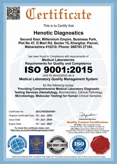
Official Diagnostic Certification Documentation
Furthermore, our official accreditation certificates demonstrate compliance with international ISO standards, atomic radiation safety guidelines, and clinical diagnostic benchmarks.



What Our Patients Say — Patient Testimonials & Clinical Experiences
Specifically, read how advanced 3.0T MRI, 128-slice CT, and ultrasound diagnostic imaging at Henotic Diagnostics Radiology Center delivered accurate clinical clarity and compassionate care for our patients.
“Specifically, I was extremely anxious about my lumbar spine MRI scan due to severe claustrophobia. However, the 3.0T wide-bore MRI system at Henotic Diagnostics Radiology Center with quiet silent scan technology made the procedure completely comfortable. Furthermore, Dr. Pratibha Patil’s detailed reporting identified my L4-L5 disc herniation within hours. Consequently, my spine specialist could start treatment immediately!”
“In addition, my referring physician advised an urgent HRCT chest scan for persistent pulmonary symptoms. The 128-slice CT scan at Henotic Diagnostics was completed in less than 5 minutes with minimal radiation. Moreover, I received the verified digital report and DICOM images directly on WhatsApp on the same evening. As a result, the diagnostic clarity gave our family huge peace of mind!”
“Additionally, we visited Henotic Diagnostics Radiology Center for my Level 2 detailed fetal anomaly scan during the second trimester. The radiologist explained every anatomical structure on the screen with immense patience and care. Furthermore, the 4D ultrasound clarity was unbelievable. Therefore, we highly recommend their PCPNDT-registered sonography suite to all expecting parents!”
“Similarly, my father required a whole-body 18F-FDG PET-CT scan for oncology treatment monitoring. The reception staff and nuclear medicine technicians treated us with wonderful empathy and courtesy throughout intake. Consequently, the high-resolution PET-CT report provided accurate tumor margin evaluation for his oncologist. Truly an exceptional radiology center!”
Frequently Asked Questions About Medical Imaging Center Services
General Medical Imaging Questions
Q: What is a Medical Imaging Center? A specialized diagnostic facility equipped with advanced radiological equipment to visual non-invasively internal organs and tissues.
Q: What are the different types of medical imaging? Core modalities include MRI, CT scan, ultrasound, Doppler, digital radiography, mammography, and PET-CT.
Q: What is the difference between imaging and a diagnostic test? Imaging captures spatial structural images of internal anatomy; lab diagnostic tests measure biochemical fluid markers.
Q: Who interprets medical imaging scans? Scans at a Medical Imaging Center are analyzed by board-certified radiologists who generate formal diagnostic reports for your physician.
Q: Are imaging tests painful? Diagnostic imaging is non-invasive and painless. Minor temporary discomfort may arise from lying still or IV needle placement.
MRI Questions
Q: Does MRI use radiation? No, 3T MRI uses strong magnetic fields and radiofrequency waves with 0 mSv ionizing radiation.
Q: Can I have an MRI with a pacemaker? Traditional pacemakers preclude MRI. Modern MR-conditional pacemakers require specific cardiology programming protocols.
Q: Why is MRI noisy? Gradient magnetic coils rapidly switching electric currents produce thumping acoustic sounds; hearing protection is provided.
Q: What if I am claustrophobic? Wide-bore 3T MRI systems, ambient lighting, and music headsets significantly reduce claustrophobic anxiety.
CT Scan Questions
Q: Does CT scan use radiation? Yes, CT scans utilize low-dose X-ray beams optimized under strict ALARA safety standards.
Q: How long does a CT scan take? Image acquisition takes seconds; the full examination process takes 5 to 15 minutes.
Q: What is HRCT Chest? High-Resolution CT uses sub-millimeter slices to evaluate lung parenchyma and interstitial lung disease.
Q: What is CT coronary angiography? A non-invasive CT scan mapping coronary arteries to detect plaque blockages without cardiac catheterization.
Ultrasound & Doppler Questions
Q: Does ultrasound use radiation? No, ultrasound utilizes high-frequency acoustic sound waves with zero radiation exposure.
Q: Do I need fasting before ultrasound? Abdominal ultrasound requires 6–8 hours fasting to prevent bowel gas and distend the gallbladder.
Q: What is Color Doppler? A specialized ultrasound measuring red blood cell velocity to map vascular blood flow in deep veins and arteries.
Mammography Questions
Q: What is 3D Mammography? Digital breast tomosynthesis acquires multi-angle images to reconstruct 3D breast tissue layers.
Q: Can mammography and ultrasound be used together? Yes, sonomammography complements digital mammography in women with dense breast tissue.
PET-CT Questions
Q: What is FDG PET-CT? A nuclear scan using injected 18F-FDG glucose radiotracer to pinpoint hyper-metabolic cancer cells.
Q: Why is fasting required before PET-CT? Fasting lowers background blood glucose so cancer cells rapidly take up the injected FDG radiotracer.
Pregnancy Imaging Questions
Q: Is ultrasound safe during pregnancy? Ultrasound does not use ionizing radiation and is widely used during pregnancy when clinically appropriate.
Q: Should I tell the Medical Imaging Center if I may be pregnant? Yes, always inform your technologist and physician prior to any scan if you are or might be pregnant.
Contrast & Safety Questions
Q: Are contrast dyes safe for everyone? Most patients tolerate contrast agents with no side effects. Patients with kidney dysfunction, asthma, or previous contrast reactions are screened prior to administration.
Q: How is radiation minimized during imaging? Scans follow strict ALARA standards, using calibrated low-dose protocols and non-ionizing alternatives (Ultrasound, MRI) wherever clinically viable.
Preparation Questions
Q: Do all medical imaging scans require fasting? Fasting is necessary for abdominal ultrasounds, contrast CT/MRI, and PET-CT. Routine X-rays and joint scans usually require no fasting.
Q: What should I wear for my imaging exam? Wear loose, comfortable clothing without metal fasteners. Remove metal jewelry, watches, and hair accessories prior to your scan.
Report & Results Questions
Q: What does “Impression” mean in a radiology report? The radiologist’s synthesized diagnostic conclusion summarizing key findings for your doctor.
Q: Who explains my scan report? Your attending physician or referring specialist is best qualified to explain your imaging report within your overall clinical care.