

Dr Pratibha Patil
MBBS, MD (Radio-Diagnosis)
16+ Years ExperienceSpecialist in: Women’s Breast Imaging, BI-RADS Classification, Breast Cancer Screening Interpretation, USG-Guided Interventions, Fetal and Obstetric Ultrasound
Connecting to our secure server
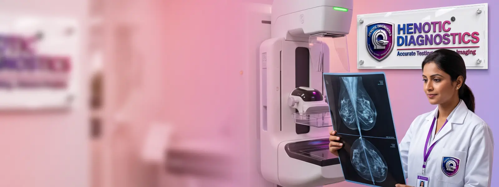
Browse our comprehensive library of diagnostic capabilities. We provide over 200 NABL accredited radiology scans and automated clinical pathology investigations.
Radiation-free Breast Cancer Screening using high-resolution bilateral Sono Mammography (Breast Ultrasound), interpreted live by senior consultant radiologists at Henotic Diagnostics, Kharghar, Navi Mumbai.

Furthermore, the table below summarises key details you need to know before booking your Breast Cancer Screening appointment at Henotic Diagnostics.
| Parameter | Details |
|---|---|
| Service | Breast Cancer Screening via Advanced Bilateral Sono Mammography |
| Modality | High-frequency Ultrasound (Sono Mammography) — no X-ray, no radiation |
| Focus | Early detection of breast cancer, cysts, fibroadenomas and lymph node abnormalities |
| Radiation Exposure | ✓ Zero Radiation |
| Duration | 20–30 minutes (bilateral scan of both breasts + axillary lymph nodes) |
| Report Turnaround | Same day — delivered digitally via WhatsApp or email |
| Report Classification | BI-RADS (0–6) by senior consultant radiologist |
| Preparation Required | No fasting; avoid deodorant or powder on breast area on the day |
| Performed By | NABL-certified senior female radiologist |
| Insurance | ✓ Accepted — Cashless Available |
| Location | Henotic Diagnostics, Kharghar, Navi Mumbai (Open 24×7) |
| Book Appointment | Call / WhatsApp: 088793 27184 |
Breast Cancer Screening is a systematic examination of breast tissue in women who have no current symptoms, with the primary goal of detecting cancer or pre-cancerous changes at the earliest possible stage. Specifically, early-stage detection dramatically improves treatment success rates and survival outcomes, as outlined in the World Health Organization (WHO) Global Breast Cancer Initiative guidelines.
At Henotic Diagnostics, the examination is performed using Advanced Bilateral Sono Mammography — a high-frequency ultrasound technique that produces real-time, high-resolution images of both breasts simultaneously. Consequently, this approach is particularly effective for women with dense breast tissue, for whom X-ray mammography may miss subtle lesions.
Additionally, Additionally, Additionally, the bilateral scan evaluates both breasts alongside the axillary lymph nodes, providing a comprehensive clinical picture for BI-RADS classification. Indeed, the procedure is conducted exclusively by NABL-certified senior female radiologists at Henotic Diagnostics, ensuring accuracy, dignity, and a fully patient-centred experience throughout the visit.

Moreover, this service is recommended for a wide range of risk factors and clinical presentations. Furthermore, early detection via the scan remains the single most effective measure to reduce breast-cancer-related mortality.
Furthermore, women with a first-degree relative diagnosed with breast cancer carry a significantly elevated lifetime risk. Consequently, annual clinical evaluation and screening are strongly advised from age 30 onwards, as detailed in the National Cancer Institute (NCI) Screening PDQ.
Moreover, women confirmed to carry BRCA1 or BRCA2 gene mutations benefit from structured surveillance protocols — typically every 6 to 12 months — established by the National Comprehensive Cancer Network (NCCN) Genetic Assessment Guidelines.
Additionally, all women aged 35 and above are advised to undergo routine Breast Cancer Screening annually, regardless of symptoms, in alignment with the American Cancer Society early detection recommendations.
Specifically, women with radiologically dense breast tissue require specifically, this examination via Sono Mammography because ultrasound detects lesions within dense tissue far more reliably than X-ray mammography alone.
Importantly, any newly discovered lump, nipple discharge, skin dimpling, or persistent breast pain warrants urgent Breast Cancer Screening to rule out malignancy and guide appropriate clinical management.
Furthermore, women who have previously undergone treatment or wish to bundle this test with comprehensive women’s health checkup packages benefit from scheduled periodic surveillance.
Conveniently, the table below compares the three principal detection modalities, drawing on clinical comparative data from the NHS Breast Screening Programme to help you choose the best diagnostic approach.
| Screening Feature | Sono Mammography (Our Method) |
Digital Mammography | Breast MRI |
|---|---|---|---|
| Radiation Exposure | ✓ Zero Radiation | ✗ Low-dose X-ray | ✓ No Radiation |
| Best for Dense Breasts | ✓ Excellent | ✗ Limited accuracy | ✓ Excellent |
| Detects Cysts vs Solid Masses | ✓ Superior | ✗ Cannot differentiate | ✓ Good |
| Breast Compression Required | ✓ No Compression | ✗ Yes — painful | ✓ None |
| Cost-Effectiveness | ✓ Affordable | ✓ Affordable | ✗ Expensive |
| Contrast Agent Required | ✓ Not Required | ✓ Not Required | ✗ Often Required |
| Real-Time Imaging | ✓ Live Scan | ✗ Static Film | ✗ Static |
| Axillary Node Evaluation | ✓ Included | ✗ Limited | ✓ Included |
| BI-RADS Report Issued | ✓ Standard | ✓ Standard | ✓ Standard |
Specifically, bilateral Sono Mammography at Henotic Diagnostics covers a comprehensive range of breast structures. The scan includes the following clinical evaluations for complete bilateral coverage in every Breast Cancer Screening visit.
| Evaluated Structure | Clinical Significance |
|---|---|
| Breast Parenchyma (both sides) | Identifies masses, asymmetry, and diffuse architectural changes early |
| Cystic Lesions | Classifies simple versus complex cysts; rules out intracystic malignancy |
| Solid Masses | Evaluates echogenicity, margins, vascularity — BI-RADS scored for Breast Cancer Screening |
| Fibrous / Fatty Tissue Ratio | Determines breast density category for risk stratification |
| Nipple–Areola Complex | Detects subareolar lesions, duct ectasia, or discharge origin |
| Axillary Lymph Nodes (bilateral) | Screens for lymph node enlargement or suspicious morphology |
| Vascularity (Colour Doppler) | Assesses blood flow to lesions — key differentiator for malignancy |
| Skin and Subcutaneous Layer | Detects skin thickening or peau d’orange changes in the breast |
| Chest Wall (superficial) | Identifies deep masses or implant-related anomalies requiring follow-up |


Moreover, the entire procedure is conducted in a private, female-only scanning suite. Subsequently, each step follows a standardised clinical protocol to ensure reliable and reproducible results.
Initially, our front-desk team registers you and reviews any previous mammogram films, biopsy reports, or earlier scan records you carry for the radiologist’s reference.
Next, Next, a trained technologist collects your family history, menstrual cycle details, hormone therapy use, and prior medical records relevant for accurate risk stratification.
Subsequently, you lie comfortably on the scan table. Subsequently, a warm water-based gel is applied to both breasts — no compression, no discomfort. The environment is designed entirely for privacy and dignity.
Then, the radiologist methodically scans each breast quadrant — upper outer, upper inner, lower outer, lower inner, and the retroareolar zone — capturing real-time images for subsequent real-time radiologist analysis.
Meanwhile, Meanwhile, both axillary regions are scanned to identify enlarged or morphologically abnormal lymph nodes, which are critical staging markers in any complete bilateral breast scan.
Following bilateral scanning, specialized Colour Doppler ultrasound evaluates blood flow within detected lesions — an essential differentiator between benign and potentially malignant masses during the examination.
Afterward, the senior radiologist reviews all images on high-definition diagnostic monitors, applies BI-RADS scoring, and annotates findings with full clinical correlation as required for accurate BI-RADS reporting.
Finally, your BI-RADS report for Breast Cancer Screening is delivered digitally the same day via WhatsApp or email, with a printed copy available for insurance claims or specialist referrals.
Indeed, your Breast Cancer Screening report is interpreted exclusively by our certified radiology and imaging specialists with extensive clinical training in breast sonology and BI-RADS reporting.
MBBS, MD (Radio-Diagnosis)
16+ Years ExperienceSpecialist in: Women’s Breast Imaging, BI-RADS Classification, Breast Cancer Screening Interpretation, USG-Guided Interventions, Fetal and Obstetric Ultrasound

MBBS, DNB (Radio-Diagnosis)
11+ Years ExperienceSpecialist in: Breast Ultrasound, Breast Cancer Screening Reporting, USG-Guided FNAC, Sono Mammography, Musculoskeletal and Vascular Ultrasound
Importantly, minimal preparation is required before the scan. However, following these simple guidelines ensures the most accurate results from your Breast Cancer Screening appointment.
| ✓ What to Do Before Your Appointment |
|---|
| Wear comfortable two-piece clothing for easy access during the scan |
| Bring all previous mammogram films, ultrasound reports, or biopsy results |
| Inform the technologist if you are pregnant or currently breastfeeding |
| Disclose any breast implants before the scan begins |
| Furthermore, bring your insurance card for cashless billing at the diagnostic centre |
| Book at least one day in advance for priority appointment scheduling |
| ✗ What to Avoid Before Your Appointment |
|---|
| Do not apply deodorant, powder or body lotion on the breast or underarm area |
| Avoid wearing underwired or padded bras on the day of your examination |
| Refrain from using strong perfume or talcum powder near the chest |
| Do not skip your appointment due to menstruation — it is safe |
| Therefore, avoid postponing your annual scan based on minor reasons |

Specifically, Henotic Diagnostics uses institutional-grade high-frequency linear array transducers calibrated for breast ultrasound, following clinical principles documented by the Radiological Society of North America (RSNA). Specifically, these advanced systems deliver spatial resolution of up to 0.1 mm — critical for detecting sub-centimetre lesions at the earliest possible stage.
Furthermore, integrated Colour Doppler and Power Doppler imaging within our workflow allow real-time perfusion mapping, enabling radiologists to characterise lesion vascularity with precision during every examination.
Additionally, our suite features Elastography — a technique that measures tissue stiffness — to distinguish soft benign cysts from stiffer potentially malignant lesions. Consequently, this reduces false-positive recall rates and improves overall ultrasound specificity.
Moreover, all reports are compliant with the standardized American College of Radiology (ACR) BI-RADS Atlas, ensuring your specialists worldwide can interpret your Breast Cancer Screening findings with full confidence.
Moreover, Henotic Diagnostics holds the highest accreditations in diagnostic radiology, ensuring that every Breast Cancer Screening examination meets international clinical quality standards.
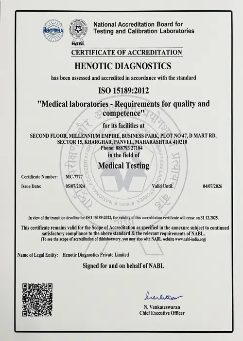 NABL Accredited
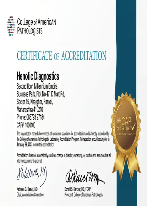 CAP Certified
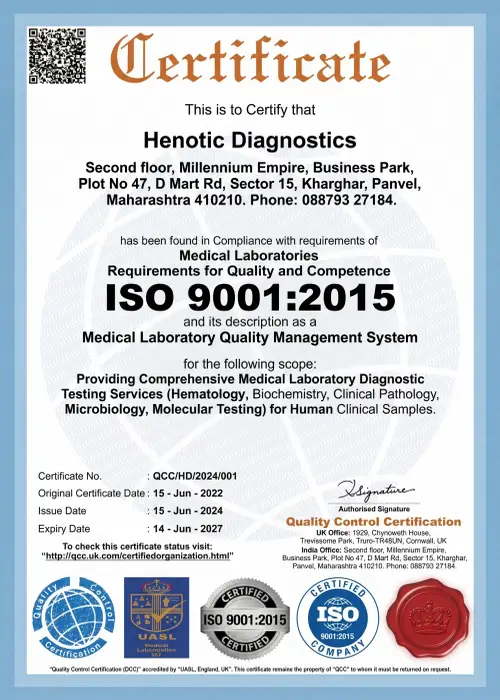 ISO 9001:2015
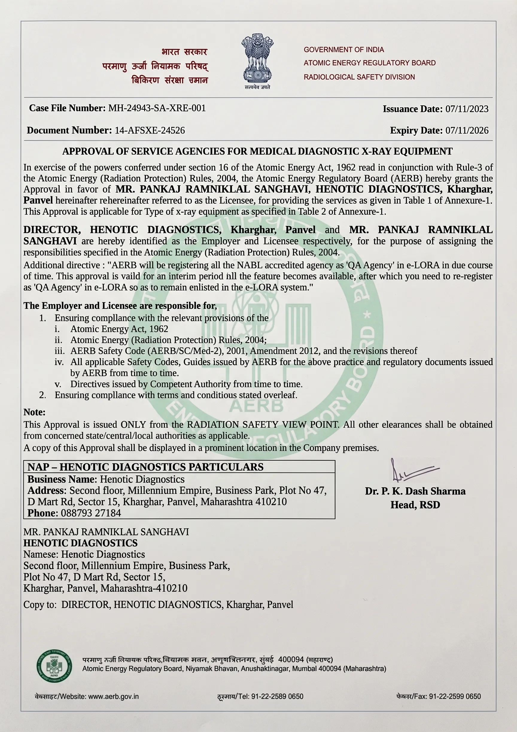 AERB Approved
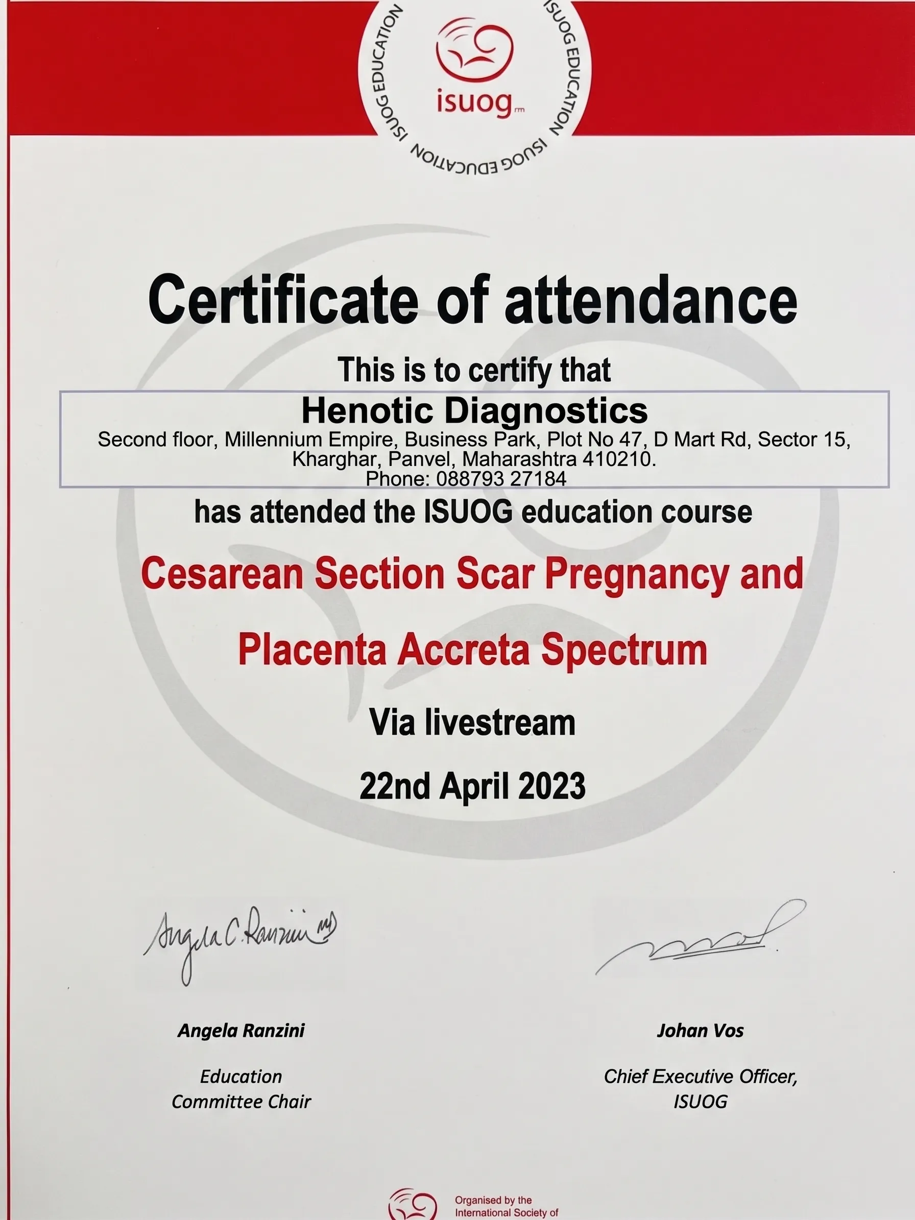 ISUOG Member
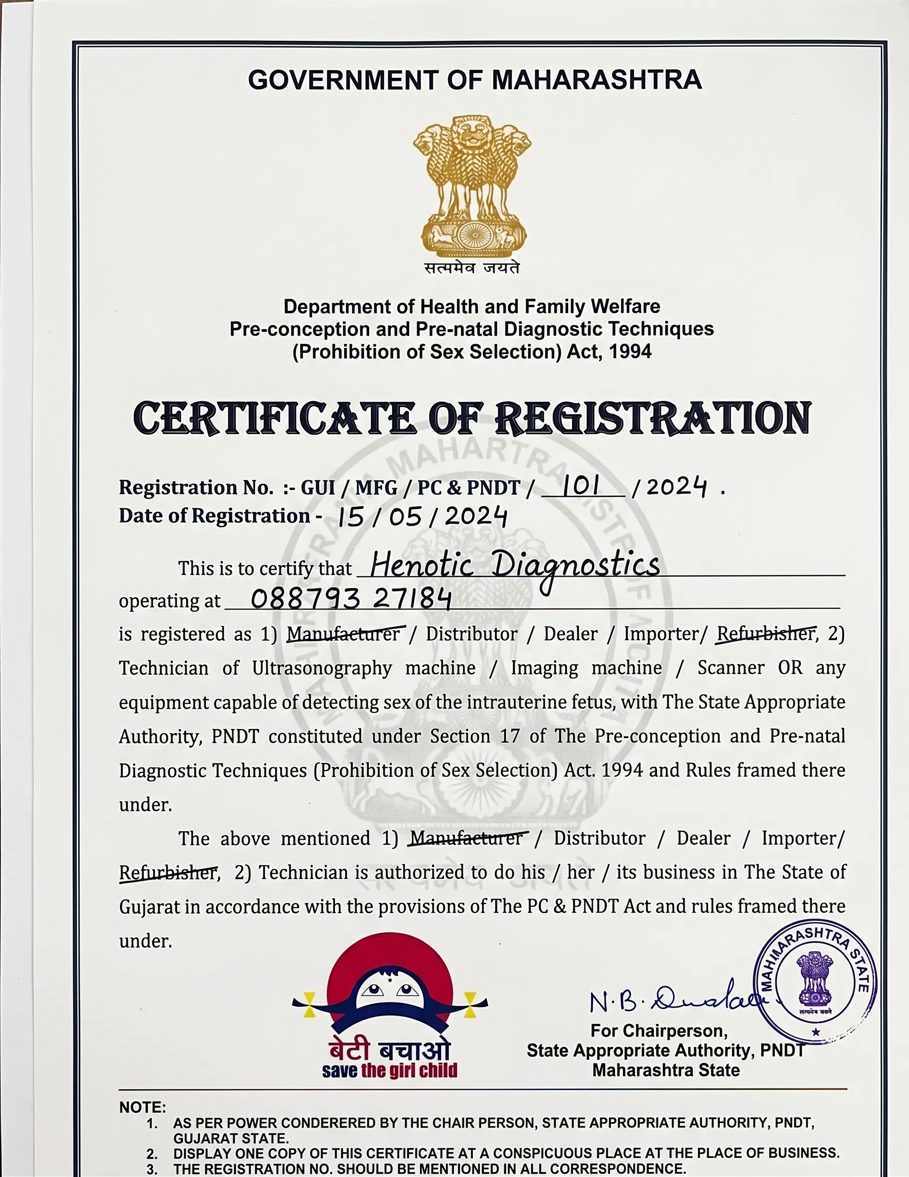 PCPNDT Registered
Additionally, our verified Google reviews reflect the compassionate, accurate, and privacy-focused experience we deliver with every consultation at Henotic Diagnostics.
“Indeed, I was extremely anxious after finding a lump. The female technician and doctor were gentle, explained everything on the screen during my Breast Cancer Screening examination, and delivered my report on WhatsApp within 2 hours. Highly recommended.”
“Moreover, the centre was very clean with polite staff. No long waiting lines. Dr. Patil reviewed my previous mammogram side-by-side with the sono mammography to confirm it was a benign cyst. Pure peace of mind — highly recommend this centre for Breast Cancer Screening near Vashi.”
“Transparent pricing with no hidden charges. Claimed my OPD insurance smoothly with their structured receipts. Consequently, I will continue annual Breast Cancer Screening here without hesitation.”
Specifically, every report at Henotic Diagnostics uses the internationally standardised BI-RADS (Breast Imaging Reporting and Data System) classification. Moreover, understanding your BI-RADS score from your Breast Cancer Screening helps you discuss findings confidently with your treating physician.
| BI-RADS Category | Finding in Breast Cancer Screening | Recommended Action | Malignancy Risk |
|---|---|---|---|
| Category 0 | Incomplete — additional imaging needed for a complete breast assessment | Supplemental mammogram or MRI | Undetermined |
| Category 1 | Negative — normal result; no suspicious findings | Annual check-up recommended | Essentially 0% |
| Category 2 | Benign finding confirmed (cysts, fibroadenoma) | Routine annual follow-up examination | 0% |
| Category 3 | Probably benign — short-term follow-up advised | Repeat Breast Cancer Screening scan in 6 months | <2% |
| Category 4 | Suspicious — biopsy recommended after the sono mammography scan | USG-guided FNAC or core needle biopsy | 2%–95% |
| Category 5 | Highly suggestive of malignancy on the scan | Biopsy + oncology referral | >95% |
| Category 6 | Known biopsy-proven malignancy — Sono Mammography for treatment planning | Oncology management plan | 100% (confirmed) |
Conveniently, Henotic Diagnostics accepts leading health insurance plans for Breast Cancer Screening. Furthermore, cashless processing is available for pre-authorised insurance policies.
| Insurance Provider | Coverage for Screening | Documents Required |
|---|---|---|
| Star Health | ✓ Covered (OPD / Day Care) | Insurance card + Photo ID + Referral letter |
| HDFC ERGO | ✓ Covered | Policy number + Referring doctor prescription |
| New India Assurance | ✓ Covered (OPD) | Policy document + Referral + ID proof |
| Niva Bupa | ✓ Covered | App e-card + Referral letter |
| Medi Assist TPA | ✓ Cashless Processing Available | TPA card + Referral |
| Ayushman Bharat / PMJAY | ✓ Covered (eligible beneficiaries) | PMJAY card + Aadhaar |
| Corporate Group Policies | ✓ Most plans — call to confirm | Employee ID + Group policy card |
Importantly, call 088793 27184 to confirm your insurer’s coverage for Breast Cancer Screening before visiting the centre.
Importantly, the following questions about Breast Cancer Screening are commonly searched. Furthermore, the accurate clinical answers below help you make a well-informed decision about your breast health.
Breast Cancer Screening is an early-detection examination that checks asymptomatic women’s breast tissue for cancer or pre-cancerous changes. At Henotic Diagnostics, the examination uses radiation-free bilateral Sono Mammography, and a BI-RADS classified report is issued on the same day by a senior radiologist.
Furthermore, women aged 35 and above, those with a family history of breast cancer, BRCA gene carriers, women with dense breast tissue, or anyone with a palpable lump or nipple discharge should prioritise annual bilateral examinations, in accordance with CDC Breast Cancer Screening guidance. Furthermore, post-treatment patients require periodic surveillance scans.
No. The procedure is completely painless and non-invasive. A smooth transducer glides gently over the breast surface with water-based gel during Breast Cancer Screening. Consequently, there is no compression and no discomfort — unlike X-ray mammography which involves breast compression.
Specifically, a complete bilateral session takes approximately 20 to 30 minutes, including real-time scanning of both breasts, axillary lymph nodes, Colour Doppler assessment, and on-the-spot radiologist review.
Breast Cancer Screening via Sono Mammography uses sound waves — no radiation, no compression. Conversely, mammography uses low-dose X-rays and involves mechanical breast compression. Specifically, the ultrasound method is more effective for dense breasts and is the preferred modality for women under 50.
The cost is affordable and completely transparent — no hidden charges. Additionally, most major health insurance plans cover Breast Cancer Screening at Henotic Diagnostics. Call or WhatsApp 088793 27184 for the current package pricing and insurance eligibility confirmation.
Furthermore, Henotic Diagnostics offers a full spectrum of NABL-certified diagnostic services alongside Breast Cancer Screening, Consequently, all services are available 24 hours a day, 7 days a week.
Conveniently, Henotic Diagnostics is located in Kharghar, Sector 15, Navi Mumbai — easily accessible from Belapur, Panvel, Vashi, Airoli and surrounding areas for sono mammography screening appointments.
Simply fill in the details to confirm your Breast Cancer Screening appointment at Henotic Diagnostics. Alternatively, you can call or WhatsApp 088793 27184 for immediate assistance and priority booking.
Get priority booking through our secure portal.