
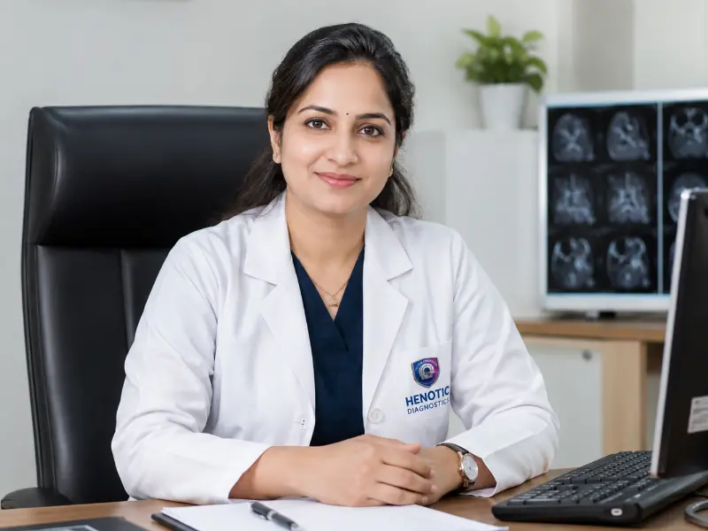
Dr. Pratibha Patil
MBBS, MD (Radio-Diagnosis)
16+ Years Experience
Specialist in Women’s Imaging, High-Resolution Breast Ultrasound, & BI-RADS Lesion Categorization.
Connecting to our secure server
Browse our comprehensive library of diagnostic capabilities. We provide over 200 NABL accredited radiology scans and automated clinical pathology investigations.
Advanced Diagnostic Radiology
Accurate, radiation-free, high-resolution breast ultrasound interpreted by certified senior consultant radiologists.

| Service Parameter | Details |
|---|---|
| Examination | Sono Mammography / Bilateral Breast Ultrasound |
| Imaging Modality | High-Frequency Linear Ultrasound (No ionizing radiation) |
| Radiation Exposure | Zero (100% Radiation-Free & Safe during pregnancy) |
| Fasting Requirement | None (Normal food and hydration permitted) |
| Preparation | Minimal (Wear comfortable, easily removable two-piece clothing) |
| Procedure Duration | Approx. 15 – 30 minutes |
| Reporting Specialists | MD / DNB Senior Consultant Radiologists (BI-RADS compliant) |
| Report Delivery | WhatsApp, Secure Email, Patient Portal & Centre Printout |
| Turnaround Time | Same-day delivery (Within 2 to 4 hours) |
| Centre Timing | Open 24 Hours / 7 Days a Week |
Sono Mammography, also known as breast ultrasound, is a non-invasive, radiation-free diagnostic imaging examination specifically calibrated to evaluate breast tissue, lumps, cysts, solid lesions, and microvascular architecture in real time.
At Henotic Diagnostics, breast ultrasound is performed using modern high-frequency linear transducers and color Doppler systems, interpreted meticulously by senior consultant radiologists adhering to international BI-RADS (Breast Imaging-Reporting and Data System) criteria.
A clinician may recommend Bilateral Sonomammography for:
While both examinations serve essential roles in breast health, they rely on distinct imaging principles and address complementary diagnostic needs.
| Evaluation Criteria | Sono Mammography (Ultrasound) | Mammography (X-Ray) |
|---|---|---|
| Underlying Technology | High-frequency sound waves | Low-dose ionizing radiation (X-rays) |
| Radiation Hazard | None (Safe for pregnancy & repeated checks) | Low-dose ionizing radiation |
| Cyst vs. Solid Mass | Gold standard: reliably distinguishes fluid from solid | Identifies density, but less definitive on internal fluid |
| Dense Breast Tissue | High sensitivity in dense glandular tissue | Sensitivity can be obscured by overlapping dense tissue |
| Microcalcifications | Limited utility for tiny microcalcifications | Superior detection of suspicious microcalcifications |
| Compression Discomfort | Gentle probe manipulation using warm gel | Firm compression of breast tissue between plates |
When a patient or doctor detects a lump, sonomammography provides instant spatial and morphological detail. The reporting radiologist analyzes:

Younger women typically exhibit dense glandular tissue that appears radiopaque (“white”) on routine mammograms, potentially masking small underlying lesions. Ultrasound penetrates dense parenchymal tissue effectively.
Because it uses zero ionizing radiation, sonomammography is the absolute first choice for examining pregnant mothers, nursing women, and patients under 35 presenting with palpable symptoms.

Accurate imaging requires experienced clinical eyes. Every study at Henotic Diagnostics is interpreted by dedicated senior diagnostic radiologists.
MBBS, MD (Radio-Diagnosis)
16+ Years Experience
Specialist in Women’s Imaging, High-Resolution Breast Ultrasound, & BI-RADS Lesion Categorization.

MBBS, DNB (Radio-Diagnosis)
11+ Years Experience
Expert in Diagnostic Breast Ultrasound, Color Doppler Studies, & USG-Guided FNAC / Interventions.
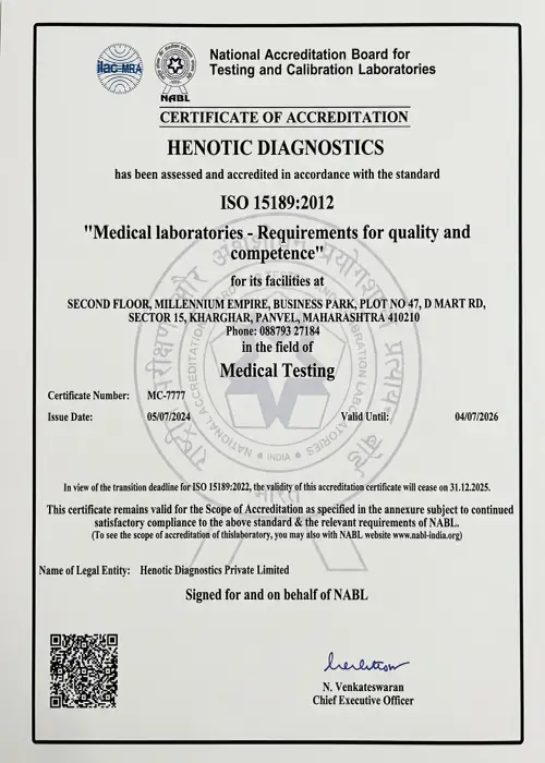
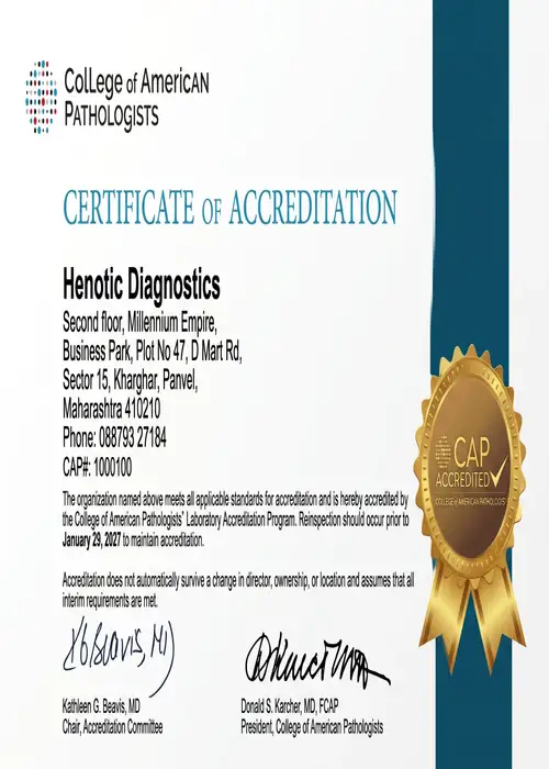
Meeting the highest national and international standards in clinical diagnostics.
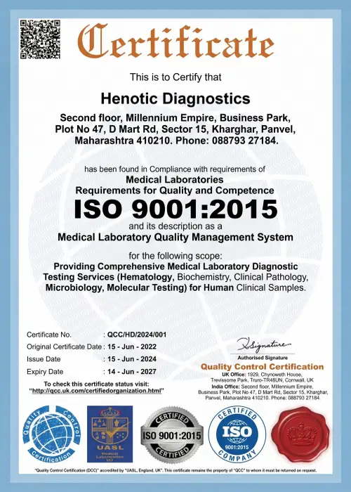

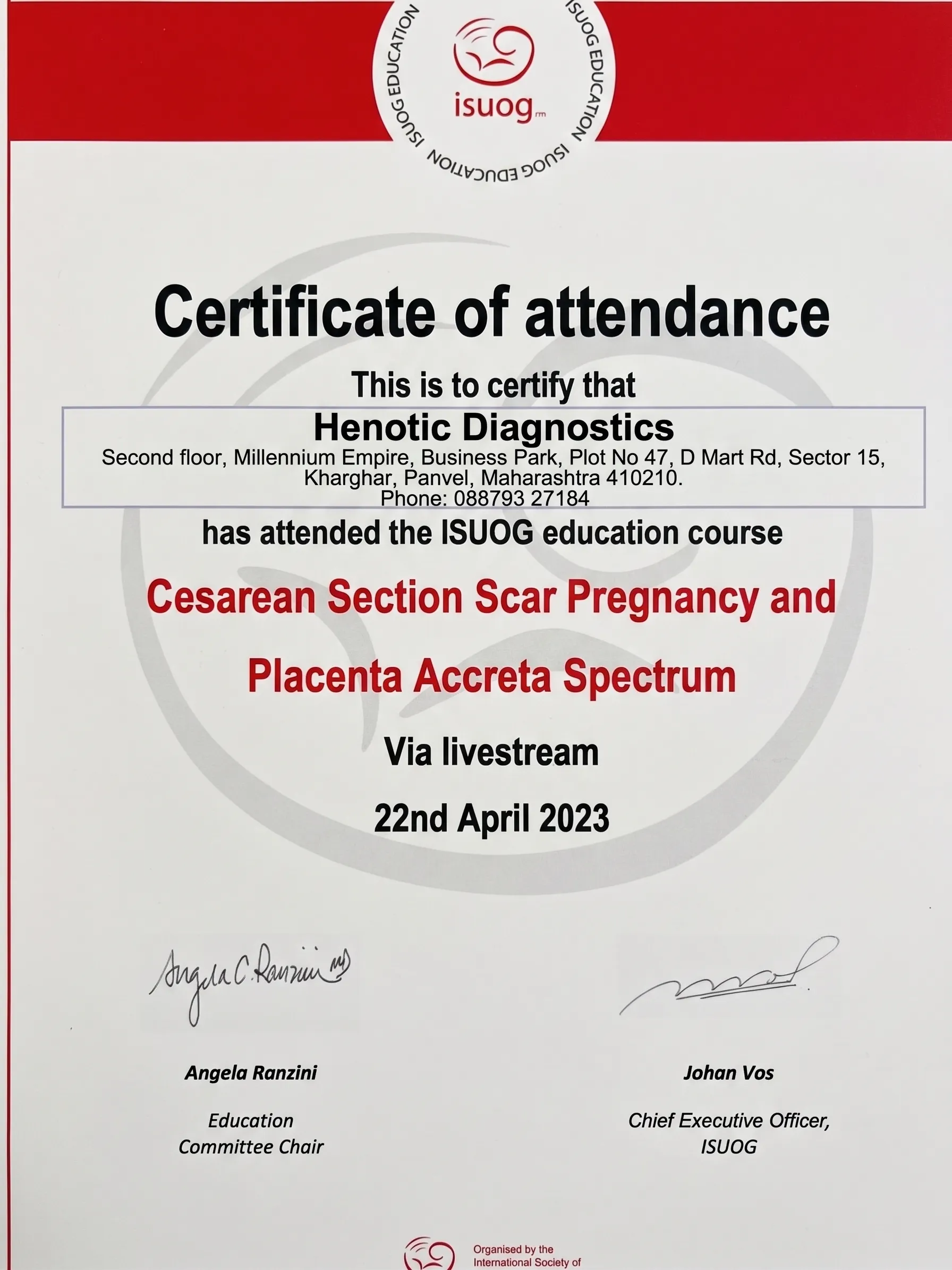
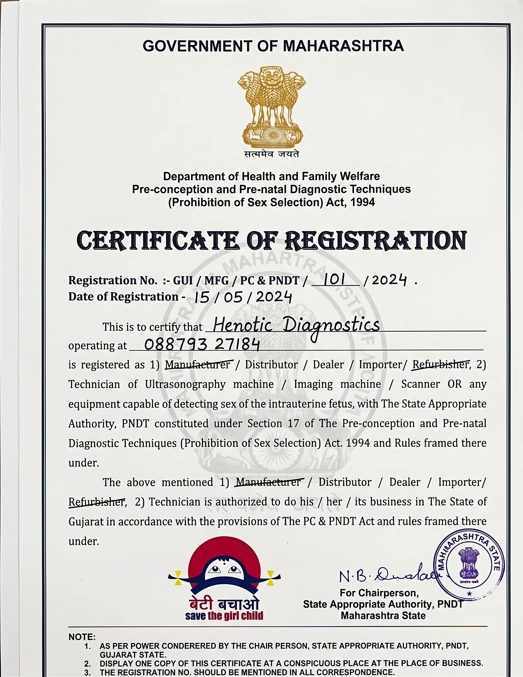
Upon check-in, patient identity is confirmed in our dedicated, private women’s imaging wing.
Our imaging team documents symptoms, prior biopsies, menstrual timing, family history, and reviews past mammograms.
You recline comfortably on the examination couch with your arm raised. Warm acoustic gel is applied to the breast surface to ensure optimal sound conduction.
The high-resolution transducer is systematically swept across all four quadrants, the retroareolar region, and the axillary lymph node beds.
Precise still frames and cinematic cine-loops of suspicious or normal structures are digitized with caliper measurements.
Our senior radiologist cross-examines findings, writes the BI-RADS assessment, and dispatches authenticated digital reports directly to your WhatsApp and email.
| Requirement | Instructions |
|---|---|
| Dietary Fasting | No fasting required. You can eat and drink normally prior to the scan. |
| Attire & Cosmetics | Wear comfortable two-piece clothing. Avoid talcum powder, deodorants, or body lotions on the chest area before your test. |
| Previous Records | Please bring past mammograms, past sonograms, FNAC/biopsy reports, or relevant imaging discs for comparative evaluation. |
| Insurance Claims | Carry your OPD insurance card/TPA claim documents if claiming diagnostic reimbursement. Structured tax invoices are issued. |
Verified feedback from patients visiting our center for breast imaging.
“I was extremely anxious after finding a lump. The female technician and doctor were gentle, explained everything on the screen, and delivered my report on WhatsApp within 2 hours. Highly recommended.”
“Very clean center with polite staff. No long waiting lines. The doctor reviewed my previous mammogram side-by-side with the sonomammography to confirm it was a benign cyst. Pure peace of mind.”
“Transparent pricing with no hidden charges. Claimed my OPD insurance smoothly with their structured receipts.”
No. Fasting is completely unnecessary for routine breast ultrasound examinations. You can maintain your normal diet and medication schedule.
No. Sonomammography relies purely on safe, high-frequency sound waves. It produces zero ionizing radiation and is entirely safe during pregnancy and lactation.
A typical bilateral scan takes between 15 to 30 minutes, allowing adequate time for a thorough evaluation of both breasts and surrounding axillary lymph nodes.
The scan is completely painless and non-invasive. You will only feel light pressure as the transducer glides over the skin with warm ultrasound gel.
BI-RADS (Breast Imaging-Reporting and Data System) is an internationally standardized score from 0 to 6 used by radiologists to classify findings, determine malignancy likelihood, and recommend specific follow-up actions.
Phone: 088793 27184
Operating Hours: Open 24 Hours (All 7 Days)
Direct Link: View on Google Maps
Get priority booking through our secure portal.