
Magnetic Resonance Medical Imaging
High-resolution soft-tissue evaluation using magnetic fields without ionizing radiation. Ideal for brain, spine, joints, and pelvic organs.
Explore MRI Services →Connecting to our secure server
Browse our comprehensive library of diagnostic capabilities. We provide over 200 NABL accredited radiology scans and automated clinical pathology investigations.
Medical Imaging refers to diagnostic techniques that create detailed images of internal anatomical structures, tissues, and organs to support clinical evaluation. At Henotic Diagnostics, our modern imaging facility is equipped with advanced technology—including 3T MRI, 128-slice CT, diagnostic ultrasound, PET-CT, digital mammography, and non-invasive cardiac diagnostics. As a result, these diagnostic scans provide detailed image resolution and qualified radiologist interpretations to assist referring clinicians in disease evaluation, diagnosis, and ongoing treatment monitoring.
In addition, our center provides comprehensive diagnostic scanning options tailored to specific anatomical areas and clinical questions. For example, patients can access high-field MRI, high-speed CT imaging, ultrasound sonography, and specialized functional studies under one roof.
High-resolution soft-tissue evaluation using magnetic fields without ionizing radiation. Ideal for brain, spine, joints, and pelvic organs.
Explore MRI Services →
Rapid cross-sectional imaging for detailed evaluation of lungs, bone fractures, acute abdominal pain, and vascular structures.
Explore CT Services →
Safe sound-wave sonography for abdominal organ assessment, pelvic evaluation, thyroid screening, and pregnancy monitoring.
Explore Ultrasound Services →
Specialized vascular ultrasound analyzing arterial and venous blood flow, deep vein thrombosis, and carotid artery circulation.
Explore Color Doppler Studies →
Low-dose digital X-rays providing rapid image processing for bone integrity, chest conditions, and joint assessment.
Explore Digital Radiography →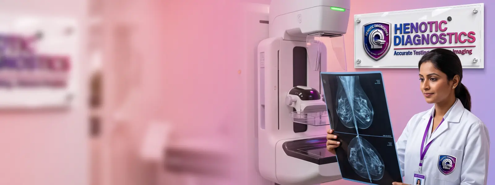
Advanced digital breast screening and diagnostic mammography designed for early microcalcification and mass detection.
Explore Mammography →
Combined anatomical and metabolic imaging providing precise tumor staging, treatment response, and cellular activity analysis.
Explore PET-CT Services →
2D Echocardiography, Stress Echo, and CT Coronary Angiography for evaluating heart valve function, ejection fraction, and arteries.
Explore Cardiac Imaging →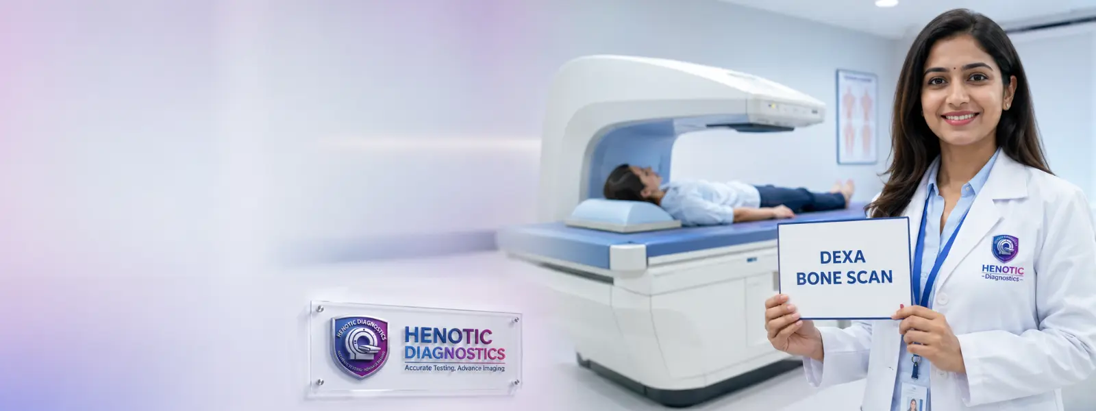
Dual-energy X-ray absorptiometry measuring bone mineral density to screen for osteoporosis and predict fracture risk.
Explore DEXA / Bone Density Imaging →
Non-invasive transient elastography assessing liver stiffness and fibrosis progression without surgical biopsy.
Explore FibroScan / Liver Elastography Imaging →Medical Imaging encompasses a set of non-invasive procedures that capture structural and functional representations of the human body. Furthermore, these techniques allow physicians to diagnose medical conditions, evaluate traumatic injuries, and monitor response to therapeutic interventions without surgical exploration.
Specifically, different modalities utilize distinct physical principles to form anatomical images. For instance, X-ray and CT scans pass controlled energy beams through tissues of varying densities, producing contrast between bone, soft tissue, and air. In contrast, Magnetic Resonance Imaging utilizes radio waves within a high-strength magnetic field to align hydrogen nuclei in cellular fluids. At the same time, ultrasound relies on high-frequency sound waves reflecting off internal tissue boundaries.
In clinical practice, medical imaging supports diagnostic evaluation across virtually all medical specialties. Consequently, referring doctors rely on these imaging modalities to evaluate:
Although laboratory blood tests measure chemical bio-markers, imaging studies provide spatial visualization of anatomical structures. Therefore, combining clinical laboratory analysis with modern medical imaging yields a far more accurate diagnostic picture for treating physicians.
Because medical technology has evolved rapidly, multiple specialized imaging modalities are currently available. Depending on the patient’s symptoms and clinical indications, physicians select the modality that provides the clearest diagnostic answers.
MRI provides detailed soft-tissue contrast without ionizing radiation. However, CT may be preferred for certain acute conditions, depending on the clinical situation. Furthermore, high-field 3T MRI systems offer enhanced spatial resolution for complex neurological imaging, joint cartilage assessment, and pelvic disease staging.
Computed Tomography creates detailed cross-sectional slices of internal organ structures using computer-assisted X-ray processing. In addition, multi-detector 128-slice CT scanners acquire volumetric data in seconds, making CT the preferred modality for emergency trauma screening, acute chest pain, and pulmonary angiography.
Diagnostic ultrasound uses non-ionizing sound waves and is widely used for fetal and pregnancy imaging when clinically indicated. Moreover, high-resolution sonography allows real-time visualization of abdominal organs, thyroid nodules, breast tissues, and superficial soft-tissue structures without thermal tissue heating or ionizing radiation.
Color Doppler ultrasound overlays directional blood flow velocity onto structural sonograms. Consequently, vascular specialists use Doppler studies to detect deep vein thrombosis (DVT), carotid artery stenosis, peripheral arterial occlusion, and placental vascular resistance.
Digital X-ray remains the foundational imaging test for primary bone fracture assessment, joint arthritis evaluation, and pulmonary infection screening. Moreover, modern flat-panel digital detectors significantly reduce exposure while producing instant digital images for immediate radiologist review.
Digital Mammography uses low-energy X-rays tailored specifically for breast tissue evaluation. Furthermore, digital breast tomosynthesis (3D mammography) captures multiple thin image slices, improving microcalcification detection in dense breast tissue.
Positron Emission Tomography combined with CT (PET-CT) merges functional metabolic data with precise anatomical cross-sections. As a result, PET-CT scans are instrumental in clinical oncology for tumor staging, monitoring treatment response, and identifying recurrent malignant lesions.
Non-invasive cardiac imaging includes 2D Echocardiography, Tissue Doppler Imaging, and Cardiac CT Angiography. Therefore, cardiologists can precisely measure ejection fraction, assess myocardial wall motion, evaluate valve regurgitation, and non-invasively visualize coronary artery narrowing.
Because each imaging modality possesses distinct technical capabilities, selecting the appropriate scan depends on the clinical question. For instance, soft tissues are best evaluated by MRI, whereas bone fractures and acute bleeding are quickly identified via CT or X-ray.
| Imaging Modality | Energy Source | Primary Clinical Indications | Key Diagnostic Strengths |
|---|---|---|---|
| MRI | Magnetic fields & RF waves | Brain, spine, joints, ligaments, pelvic organs | Superior soft-tissue contrast; no ionizing radiation |
| CT Scan | Ionizing X-rays | Trauma, lungs, acute abdomen, vascular angio | Rapid scan speed; excellent bone and calcification detail |
| Ultrasound | High-frequency sound waves | Abdomen, pelvis, pregnancy, thyroid, blood flow | Real-time imaging; highly safe for obstetrics; no ionizing radiation |
| Digital X-Ray | Ionizing X-rays | Bone fractures, chest infections, joint space | Widely accessible, fast, low-cost screening tool |
| PET-CT | Radiotracers & CT X-rays | Oncology staging, metabolic tumor mapping | Combines metabolic cellular data with anatomical imaging |
While MRI excels at detailing soft tissues such as brain parenchyma, spinal discs, and knee ligaments, CT scans offer unmatched speed for acute trauma and superior resolution for bony structures. Therefore, emergency departments often utilize CT first for head trauma, whereas outpatient neurologists prefer MRI for chronic headaches.
Both MRI and ultrasound operate without ionizing radiation. However, ultrasound is portable and provides dynamic, real-time imaging of moving structures like blood vessels and developing fetuses. In contrast, MRI provides comprehensive multi-planar deep tissue visualization inside joint capsules and pelvic cavities.
Although both modalities use X-rays, a standard digital X-ray produces a single two-dimensional projection. Conversely, a CT scan reconstructs hundreds of three-dimensional cross-sectional slices, allowing radiologists to inspect internal organ parenchymal details without tissue overlap.
Ultrasound is the initial diagnostic modality for gallbladder stones and pelvic pain due to its lack of radiation and high soft-tissue sensitivity. However, when bowel gas obscures abdominal visualization, CT scanning provides unobstructed clarity across the entire retroperitoneum.
Henotic Diagnostics performs targeted diagnostic scanning across all anatomical regions. In addition, specialized protocol parameters are tailored to each organ system to ensure optimal clinical diagnostic sensitivity.
MRI brain for stroke, tumors, multiple sclerosis; CT head for acute intracranial hemorrhage and skull fractures.
MRI cervical/lumbar spine for disc herniation and nerve compression; CT spine for complex vertebral fracture mapping.
High-Resolution CT (HRCT) chest for interstitial lung disease; digital chest X-ray for pneumonia and pleural effusion screening.
Ultrasound for gallstones and fatty liver; contrast CT abdomen for appendicitis, diverticulitis, and organ masses.
Pelvic ultrasound and dedicated multi-parametric MRI for uterine fibroids, ovarian cysts, and prostate disease evaluation.
3D Mammography combined with high-frequency breast ultrasound for comprehensive dense tissue breast lesion evaluation.
2D Echocardiography and CT Coronary Angiography for evaluating coronary plaque burden and valvular function.
High-field MRI for ACL tears, rotator cuff injuries, and meniscus lesions; digital X-ray for joint space narrowing.
Color Doppler ultrasound and CT Angiography assessing carotid stenosis, DVT, peripheral vascular disease, and aneurysms.
Diagnostic imaging services support referring doctors across major clinical medical specialties. As a result, structured diagnostic reports are customized to satisfy specific clinical criteria.
High-field MRI and neuro-CT assist neurologists in diagnosing ischemic stroke, demyelinating diseases, brain aneurysms, and neurodegenerative disorders.
Musculoskeletal radiologists evaluate tendon tears, bone fractures, osteomyelitis, and cartilage degeneration using high-resolution MRI, digital radiography, and CT reconstructions.
Non-invasive cardiac imaging techniques allow cardiologists to accurately measure ejection fraction, assess wall motion abnormalities, and screen for coronary artery blockages.
PET-CT, multi-slice CT, and contrast MRI enable oncologists to detect primary malignancies, map regional lymph node involvement, and monitor response to chemotherapy.
Dedicated women’s imaging encompasses 3D mammography, pelvic sonography, follicular tracking, and specialized breast MRI for high-risk screening.
Abdominal CT, MRCP (magnetic resonance cholangiopancreatography), and FibroScan help gastroenterologists evaluate biliary obstruction, chronic pancreatitis, and liver cirrhosis.
Obstetric ultrasound utilizes non-ionizing sound waves for routine fetal anomaly scans, growth assessment, biophysical profiles, and umbilical Doppler studies.
In certain clinical cases, contrast media (iodinated contrast for CT or gadolinium for MRI) is administered intravenously. Consequently, contrast agents highlight vascular structures and tissue perfusion, helping radiologists differentiate benign lesions from active inflammation or tumors.
Patient safety is fundamental across all diagnostic imaging protocols at Henotic Diagnostics. Therefore, our facility strictly adheres to radiation reduction guidelines, equipment calibration standards, and proper screening procedures.
Both MRI and Ultrasound modalities operate without ionizing radiation. Thus, they are inherently suitable for repeat follow-up examinations and pediatric imaging when clinically indicated.
CT scans, Digital X-rays, and Mammography utilize controlled ionizing X-rays. However, Henotic Diagnostics employs low-dose protocol settings and automatic exposure control to minimize radiation exposure while maintaining diagnostic image sharpness.
PET-CT scans involve minor radiotracer injections that decay rapidly within hours. In practice, patients are advised to hydrate post-scan to accelerate natural radiotracer clearance.
Diagnostic ultrasound is the primary modality used during pregnancy because sound waves pose no radiation risks to the fetus. Conversely, CT scans involving the abdominal or pelvic region are generally avoided during pregnancy unless emergency clinical circumstances mandate diagnostic imaging evaluation.
Preparation requirements vary depending on the ordered scan type. However, adhering to pre-scan instructions ensures high image clarity and smooth testing procedures.
Because MRI machines contain powerful magnets, patients must remove all metallic objects, jewelry, and metal accessories. In addition, patients with pacemakers, metallic implants, or aneurysm clips must inform staff beforehand for safety screening.
For contrast-enhanced CT scans, patients may need to fast for 4 to 6 hours prior to the appointment. Furthermore, serum creatinine blood testing is checked before contrast administration to verify renal function.
Abdominal ultrasound scans generally require 6 hours of fasting to decrease bowel gas and ensure gallbladder distension. On the other hand, pelvic and pregnancy ultrasounds require a full urinary bladder for optimal acoustic window visualization.
Standard digital X-rays require minimal preparation. However, patients are asked to wear comfortable clothing without metal zippers or buttons over the scanned region.
Following scan completion, images are processed through high-resolution PACS systems. Qualified radiologists analyze the diagnostic scans and issue detailed digital reports accessible online for referring clinicians and patients.
Henotic Diagnostics combines advanced imaging technology, qualified medical experts, and patient-centric care to deliver precise diagnostic insights.
Comprehensive imaging options including 3T MRI, 128-slice CT, Ultrasound, Doppler, Digital X-Ray, and PET-CT housed in one center.
Diagnostic images are systematically interpreted by qualified radiologist specialists experienced in sub-specialty clinical reporting.
Adherence to statutory radiation safety and laboratory quality standards ensures consistent diagnostic reliability.
Instant digital image transfer, cloud PACS access, and rapid report turnaround times support urgent clinical decision-making.
Specifically, expert diagnostic imaging interpretation at Henotic Diagnostics Radiology Center is led by our distinguished consultant radiologist.

Reporting Radiologist
Over 15 years of dedicated sub-specialty experience in diagnostic cross-sectional imaging, MRI interpretation, and ultrasound sonography.
Dr. Pratibha Patil oversees the radiology diagnostic reporting workflow at Henotic Diagnostics. Furthermore, her clinical expertise encompasses neuro-radiology MRI, body CT angiography, pelvic sonography, and multi-modality cancer imaging.
By working closely with referring clinicians, Dr. Patil ensures that every imaging examination yields clear, actionable diagnostic findings to guide timely patient care.
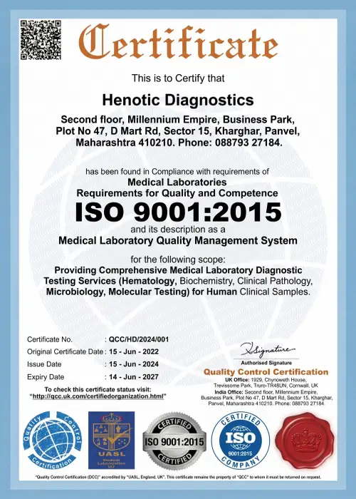
Medical Imaging Quality and Safety standards at Henotic Diagnostics strictly align with national statutory regulatory authorities and international quality frameworks.
.webp)
Radiation Safety & Diagnostic Equipment Quality Assurance

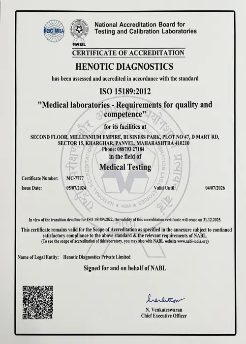
Laboratory Quality & Diagnostic Competency Accreditation

International Quality Management System Certified

International Society of Ultrasound in Obstetrics & Gynecology

Healthcare Facility Quality & Patient Safety Protocols

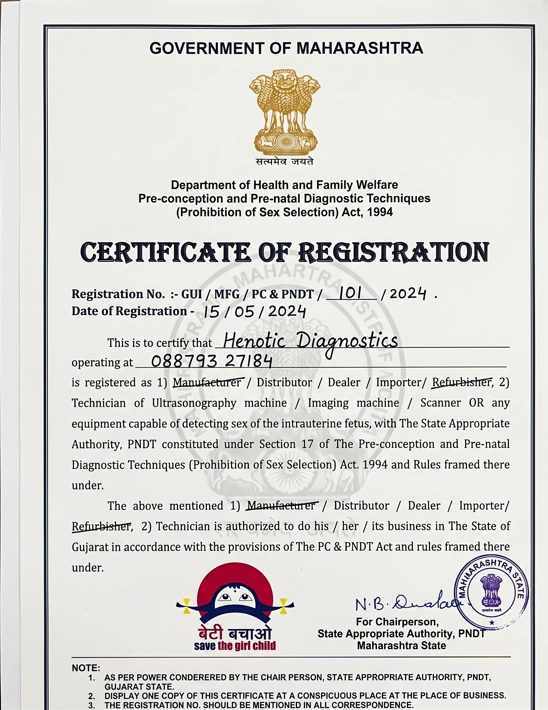
Full Compliance with Pre-Conception & Pre-Natal Diagnostic Techniques Act


Specifically, hear how our medical imaging team supports patients through clear communication, efficient scheduling, and precise diagnostic reporting.
“I underwent an MRI spine scan at Henotic Diagnostics. Furthermore, the MRI room was clean, the staff explained the procedure clearly, and the digital report was delivered to my doctor promptly.”
— Rajesh M., Kharghar
“My doctor requested an abdominal ultrasound and Doppler study. In addition, the sonographer was gentle, efficient, and took time to ensure my comfort throughout the examination.”
— Sunita S., Navi Mumbai
“The CT scan procedure was fast and well-coordinated. Consequently, receiving digital report access on my phone saved me an extra trip to collect physical films.”
— Amit K., Panvel
Henotic Diagnostics is conveniently situated in Sector 15, Kharghar, offering easy access and dedicated booking support for all medical imaging appointments.
Address: Second floor, Millennium Empire, Business Park, Plot No 47, D Mart Rd, Sector 15, Kharghar, Navi Mumbai, Panvel, Maharashtra 410210
Phone / Booking Helpline: +918879327184
24 Hours Open
View Location on Google Maps Book Scan AppointmentHere are answers to common questions regarding diagnostic imaging scans, safety considerations, preparation, and reporting procedures.
Medical imaging refers to diagnostic techniques used to create visual representations of internal body structures. For example, these techniques include MRI, CT, ultrasound, X-ray, and PET-CT, allowing clinicians to evaluate organs, bones, and tissues non-invasively.
The primary modalities include Magnetic Resonance Imaging (MRI), Computed Tomography (CT), Diagnostic Ultrasound, Digital Radiography (X-Ray), Mammography, Color Doppler, PET-CT, and Echocardiography.
No. MRI operates using strong magnetic fields and radiofrequency pulses. Therefore, it does not expose patients to ionizing radiation.
MRI provides detailed soft-tissue contrast without ionizing radiation. However, CT uses X-rays to acquire fast cross-sectional images, making CT preferred for acute trauma, bone fractures, and chest conditions.
Yes. Diagnostic ultrasound uses non-ionizing sound waves and is widely used for fetal and pregnancy imaging when clinically indicated.
Yes. CT scans utilize ionizing X-rays. However, Henotic Diagnostics employs low-dose scan protocols to minimize radiation exposure while maintaining high diagnostic clarity.
Contrast agents are administered when doctors need to highlight vascular structures, tissue perfusion, or tumor boundaries. Renal function tests are performed prior to contrast administration for patient safety.
Preparation depends on the modality. For instance, MRI requires removing metallic items, CT contrast scans may require fasting, and abdominal ultrasounds usually require 6 hours of fasting.
Medical imaging scans are interpreted by qualified radiologists who prepare diagnostic reports for referring clinicians.
The ‘Impression’ section provides the radiologist’s overall diagnostic conclusion summarizing the significant findings observed across the examination images.
Explore our complete range of diagnostic imaging services and specialized scans at Henotic Diagnostics:
Get priority booking through our secure portal.
