Advanced Medical Imaging: Types, Tests, Scans & Diagnostic Services
Medical Imaging is an essential cornerstone of modern healthcare, encompassing advanced diagnostic procedures that produce high-resolution visual representations of internal body structures, organs, and tissues for clinical evaluation.
Additionally, at Henotic Diagnostics, our modern diagnostic center offers comprehensive 3T MRI, 128-slice CT, diagnostic ultrasound, PET-CT, digital mammography, and non-invasive cardiac scans. As a result, these Medical Imaging diagnostic studies provide exceptional image clarity and expert radiologist findings to support accurate clinical diagnosis and treatment planning.
learn About medical imaging scans by Body Region
Furthermore, click on any anatomical region to navigate directly to targeted diagnostic scanning protocols:
interactive Modality & Symptom Finder
Select your symptom or body region to instantly view the clinically recommended diagnostic scan type and rationale:
3T Lumbar / Cervical spine MRI Medical Imaging
Clinical Rationale: 3T MRI is the gold-standard modality for evaluating spinal disc herniation, nerve root compression, spinal cord parenchyma, and soft-tissue ligamentous strain.
3T Neuro MRI Medical Imaging & Brain Angiogram (or CT Head for Acute Trauma)
Clinical Rationale: MRI Brain delivers multi-planar anatomical slices for evaluating ischemic stroke, demyelinating plaques (MS), tumors, and aneurysms. Moreover, for acute head trauma, CT head is preferred for immediate fracture & hemorrhage mapping.
3T High-Field Musculoskeletal (MSK) MRI Medical Imaging
Clinical Rationale: High-field 3T MRI captures sub-millimeter joint cartilage resolution, ACL/PCL ligament tears, meniscus lesions, and bone marrow edema not visible on standard X-rays.
abdominal Diagnostic Ultrasound Medical Imaging (or Contrast CT Abdomen)
Clinical Rationale: Ultrasound is the first-line non-ionizing scan for gallstones, fatty liver, renal calculi, and pelvic pain. Specifically, contrast CT abdomen provides deep retroperitoneal clarity for acute appendicitis or bowel pathologies.
high-Resolution CT medical imaging scans (HRCT) Chest / Digital chest X-Ray
Consequently, Clinical Rationale: High-Resolution CT (HRCT) provides sub-millimeter (0.35mm) cross-sectional lung parenchyma detail for acute chest pain, pulmonary embolism, or pneumonia. As a result, digital X-ray provides rapid primary chest screening.
digital 3D breast mammography Medical Imaging (Tomosynthesis) + Breast US
Therefore, Clinical Rationale: 3D Mammography (Tomosynthesis) captures thin cross-sectional breast tissue projections to isolate microcalcifications in dense tissue, paired with ultrasound for cyst differentiation.
medical Imaging Services at Henotic Diagnostics
Our center provides comprehensive diagnostic scanning options tailored to specific anatomical areas and clinical questions. Patients can access high-field MRI, high-speed CT imaging, ultrasound sonography, and specialized functional studies under one roof.
magnetic Resonance Medical Imaging
Magnetic Resonance Imaging provides high-resolution soft-tissue evaluation using magnetic fields without ionizing radiation for neurological evaluation, joint assessment, and detailed study of pelvic organs.
Find MRI Services →
Computed Tomography Medical Imaging
High-speed cross-sectional X-ray imaging provides detailed slices of bone trauma, lung parenchyma, acute abdomen, and vascular structures.
Inspect CT Services →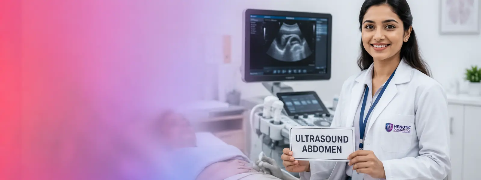
diagnostic Ultrasound Medical Imaging
High-frequency sound wave imaging provides real-time diagnostic evaluation for abdominal organs, pelvic structures, thyroid screening, and pregnancy monitoring.
See Ultrasound Services →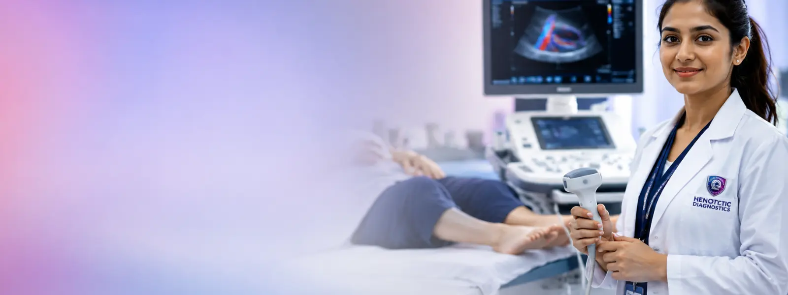
color Doppler Medical Imaging Scans
Specialized vascular ultrasound analyzing arterial and venous blood flow, deep vein thrombosis, and carotid artery circulation.
Check Color Doppler Studies →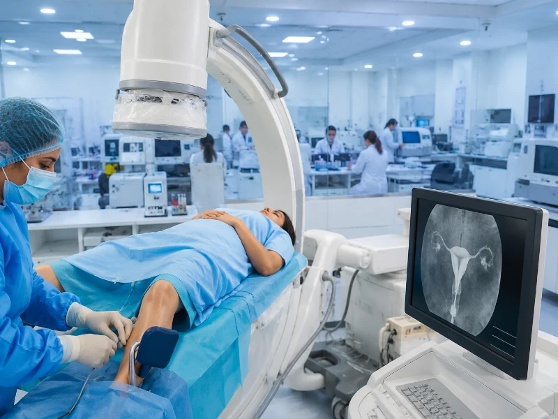
digital Radiography Medical Imaging
Low-dose digital X-rays providing rapid image processing for bone integrity, chest conditions, and joint assessment.
Examine Digital Radiography →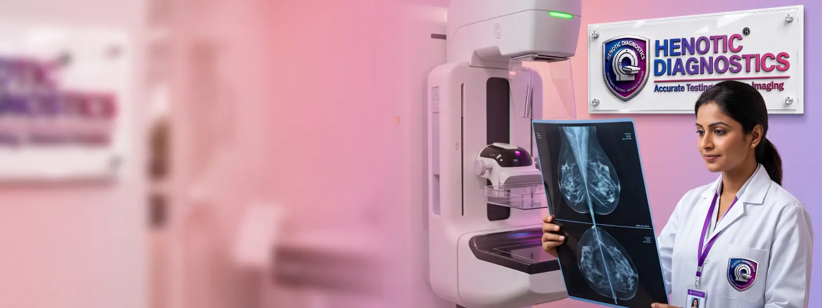
Digital Mammography Medical Imaging
Advanced digital breast screening and diagnostic mammography designed for early microcalcification and mass detection.
Access Mammography →
pET-CT & Nuclear Medicine Medical Imaging
Combined anatomical and metabolic imaging providing precise tumor staging, treatment response, and cellular activity analysis.
View PET-CT Services →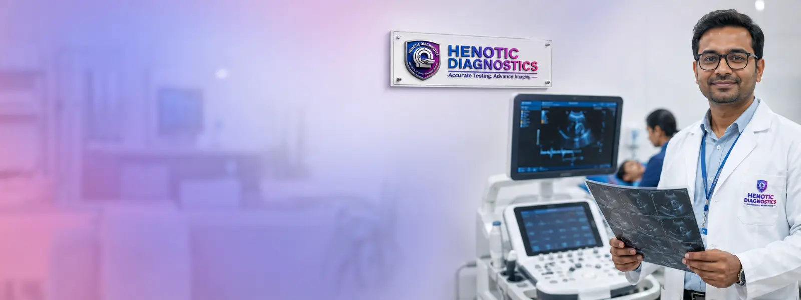
non-Invasive Cardiac Imaging — Medical Imaging Services
2D Echocardiography, stress echo, and CT Coronary Angiography for evaluating heart valve function, ejection fraction, and arteries.
Inspect Cardiac Imaging Options →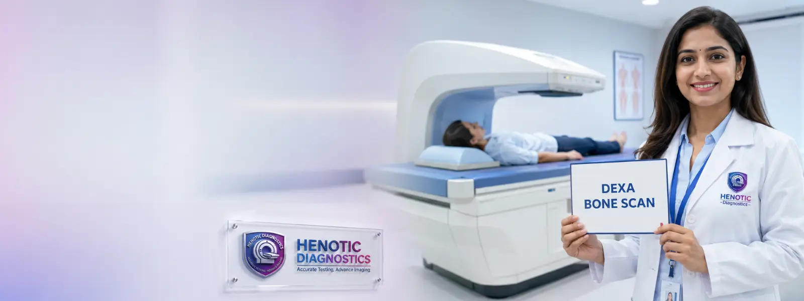
dEXA Medical Imaging Scans / Bone Density Imaging
Dual-energy X-ray absorptiometry measuring bone mineral density to screen for osteoporosis and predict fracture risk.
Discover DEXA / Bone Density Imaging →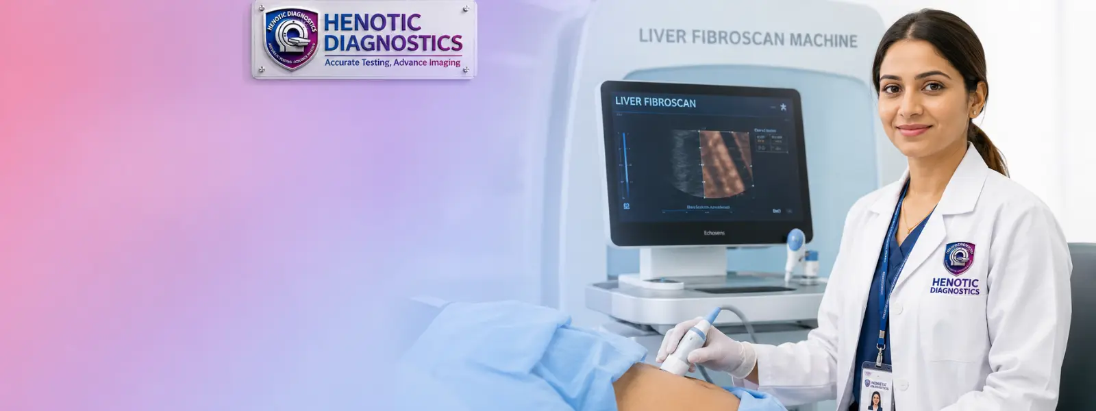
fibroScan Medical Imaging / liver elastography
Non-invasive transient elastography assessing liver stiffness and fibrosis progression without surgical biopsy.
Check FibroScan / Liver Elastography Imaging →advanced Medical Imaging by Anatomical Body Part
In fact, henotic Diagnostics performs targeted diagnostic scanning across all anatomical regions. Notably, specialized protocol parameters are tailored to each organ system to ensure optimal clinical diagnostic sensitivity.
Brain — Medical Imaging Services
Likewise, MRI brain protocols evaluate stroke, tumors, and demyelination. Specifically, hyperacute stroke protocols evaluate DWI-Perfusion Mismatch, Hyperdense MCA Sign, Time-of-Flight (TOF) Collaterals, Penumbra-to-Core Ratio, and ASPECTS Score (0-10) for emergency thrombolysis. Furthermore, ASL and Dynamic Susceptibility Contrast (DSC) Perfusion quantify blood flow, MR Spectroscopy (Choline/NAA Ratio) and 18F-FET PET Scan differentiate Pseudoprogression vs True Progression using RANO Criteria, while Phase-Contrast Velocity MRI assesses Transverse Sinus Stenosis, Optic Nerve Sheath Diameter (ONSD), Empty Sella Turcica, and Aqueductal Stroke Volume in hydrocephalus and IIH, with NI-RADS (Head & Neck) guiding oncologic surveillance.
Spine — Medical Imaging Scans Services
To clarify, multi-planar spine MRI evaluates intervertebral disc herniation and spinal stenosis. Moreover, quantitative Pfirrmann Disc Grading and Modic Endplate Changes characterization assess progressive disc degeneration, supported by multi-detector CT spine trauma mapping.
Chest & Lungs — Medical Imaging Services
High-Resolution CT (HRCT) chest for interstitial lung disease; digital chest X-ray for pneumonia and pleural effusion screening.
Abdomen — Diagnostic Imaging Services
Furthermore, diagnostic abdominal ultrasound evaluates gallstones, while contrast CT and MR Enterography assess bowel inflammation. Additionally, MRCP (MR Cholangiopancreatography) and 3T MRI with Hepatobiliary Contrast (Gadoxetic Acid) deliver focal liver lesion staging using LI-RADS® v2018. Moreover, Secretin-Enhanced MRCP evaluates Intraductal Papillary Mucinous Neoplasm (IPMN), Main Pancreatic Duct (MPD) Caliber, Pancreatic Intraepithelial Neoplasia (PanIN), and Parenchymal Atrophy Index, while Bosniak v2019 Classification, Contrast Septal Enhancement, Sub-Centimeter Renal Mass Protocol, Dual-Phase CT Urogram, and MR Urography Non-Contrast characterize complex renal cysts and urothelial masses.
Pelvis — Medical Imaging Services
Moreover, Pelvic Ultrasound and Multi-Planar T2 High-Resolution Pelvic MRI map Deep Infiltrating Endometriosis (DIE), Enzian Classification Score, Uterosacral Ligament Nodularity, and Rectovaginal Septum Lesion extension. Furthermore, prostate MRI utilizes Ultra-High B-Value Diffusion (b=2000), ADC Map Quantitative Cutoff, DCE Focal Early Washout, and PIRADS® 4/5 Index Lesion mapping for targeted In-Bore MRI Fusion Biopsy. Additionally, per ISUOG Guidelines, obstetric scans include Detailed Anomaly Scan (Level-II), Targeted Fetal Echocardiography, Uterine Artery Doppler Waveform analysis, and Fetal Biometry Percentiles tracking.
Breast — Imaging Examinations Services
Specifically, 3D Mammography combined with high-frequency breast ultrasound enables comprehensive dense tissue breast lesion evaluation. Furthermore, Contrast-Enhanced Spectral Mammography (CESM) and High-Risk Breast MRI Screening analyze Background Parenchymal Enhancement (BPE) and Ultrafast Kinetic MRI curves for precise BI-RADS® 6 Tracking and surgical monitoring.
Heart — Medical Imaging Scans Services
Additionally, 2D Echocardiography and CT Coronary Angiography enable evaluation of coronary plaque burden, valvular function, and Agatston Calcium Percentile. Specifically, FFR-CT hemodynamics and CAD-RADS™ 2.0 standardized reporting identify High-Risk Plaque (Positive Remodeling). Furthermore, Myocardial Extracellular Volume (ECV), Parametric T1/T2 Mapping, Lake Louise Criteria (Myocarditis), Amyloidosis Subendocardial Retention, and Transmural Scar Burden provide non-invasive cardiac tissue characterization alongside Myocardial Perfusion SPECT, Rubidium-82 PET Stress Scan, Fluorodeoxyglucose (FDG) Viability Study, Coronary Flow Reserve (CFR), and Transient Ischemic Dilation (TID) ischemia metrics.
Musculoskeletal & Joints — Radiology Studies Services
As a result, high-field MRI delivers detailed evaluation of ACL tears and rotator cuff injury through quantitative T2 Cartilage Mapping. Furthermore, MARS and Metal Artifact Suppression DECT eliminate metal artifacts, while Dual-Energy CT (DECT) utilizing Material Decomposition and Virtual Monochromatic Imaging (VMI) enables Urate Crystal Mapping (Gout). Additionally, Dynamic MSK Sonography, Shear-Wave Elastography (SWE), Tissue Stiffness Kilopascals (kPa), Microvascular Power Doppler, and High-Resolution Nerve Hydrodissection assess Enthesopathy & Tendinopathy, Achilles Tendon Micro-Tear, Myofascial Trigger Point Stiffness, Quantitative Fibrosis Score, and Subacromial Impingement Dynamic Test motion alongside dual-energy DEXA WHO T-Score / Z-Score bone density and Body Composition DEXA metrics.
Vascular System — Medical Imaging Services
Therefore, duplex Color Doppler ultrasound and CT Angiography assess carotid stenosis, deep vein thrombosis, peripheral arterial disease, and abdominal aortic aneurysms. Specifically, quantitative hemodynamic spectral analysis measures Peak Systolic Velocity (PSV), End-Diastolic Velocity (EDV), Resistive Index (RI), and non-invasive Ankle-Brachial Index (ABI) ratios to quantify arterial stenosis severity.
advanced Medical Imaging Services for Clinical Specialties
For example, diagnostic imaging services support referring doctors across major clinical medical specialties. Specifically, interventional radiology procedures utilize CT-Guided Biopsy / Drainage, Ultrasound-Guided FNAC, Fluoroscopic Vascular Mapping, Cone-Beam CT (CBCT), and 3D Needle Trajectory Path Planning. Furthermore, endocrine neck ultrasound applies ACR TI-RADS™ Point Score criteria, Punctate Echogenic Foci evaluation, Microcalcification Margin Analysis, and High-Risk Cervical Lymphadenopathy mapping for precise 22-Gauge Ultrasound-Guided FNAC targeting.
neurology — Medical Imaging Services
High-field MRI and neuro-CT assist neurologists in diagnosing ischemic stroke, demyelinating diseases, brain aneurysms, and neurodegenerative disorders.
orthopedics — Medical Imaging Scans Services
Musculoskeletal radiologists evaluate tendon tears, bone fractures, osteomyelitis, and cartilage degeneration using high-resolution MRI, digital radiography, and CT reconstructions.
cardiology — Medical Imaging Services
Non-invasive cardiac imaging techniques allow cardiologists to accurately measure ejection fraction, assess wall motion abnormalities, and screen for coronary artery blockages.
oncology — Medical Imaging Services
PET-CT, multi-slice CT, and contrast MRI enable oncologists to detect primary malignancies, map regional lymph node involvement, and monitor response to chemotherapy.
women’s Imaging — Medical Imaging Services
Dedicated women’s imaging encompasses 3D mammography, pelvic sonography, follicular tracking, and specialized breast MRI for high-risk screening.
gastroenterology — Medical Imaging Services
Abdominal CT, MRCP (magnetic resonance cholangiopancreatography), and FibroScan help gastroenterologists evaluate biliary obstruction, chronic pancreatitis, and liver cirrhosis.
fetal / Pregnancy Imaging — Medical Imaging Scans Services
Obstetric ultrasound utilizes non-ionizing sound waves for routine fetal anomaly scans, growth assessment, biophysical profiles, and umbilical Doppler studies.
visual Resolution: Standard 1.5T vs. In particular, henotic 3T MRI Studies
Drag the interactive slider below to see how 3T High-Field MRI delivers sub-millimeter tissue clarity for micro-cartilage tears and nerve pathways:
3T MRI (Sub-Millimeter High Field)
Standard 1.5T MRI
choosing the Right Medical Imaging Test for Clinical Needs
Furthermore, because each imaging modality possesses distinct technical capabilities, selecting the appropriate scan depends on the clinical question. Soft tissues are best evaluated by MRI, whereas bone fractures and acute bleeding are quickly identified via CT or X-ray.
comparing Medical Imaging Modalities: MRI vs CT vs Ultrasound vs X-Ray
| For this reason, imaging Modality | Besides, energy Source | However, primary Clinical Indications | Likewise, key Diagnostic Strengths |
|---|---|---|---|
| MRI | For this reason, magnetic fields & RF waves | In addition, brain, spine, joints, ligaments, pelvic organs | Furthermore, Superior soft-tissue contrast; no ionizing radiation |
| In comparison, CT Scan | Moreover, ionizing X-rays | Specifically, trauma, lungs, acute abdomen, vascular angio | Rapid scan speed; excellent bone and calcification detail |
| Additionally, Ultrasound | Consequently, high-frequency sound waves | As a result, abdomen, pelvis, pregnancy, thyroid, blood flow | Therefore, Real-time imaging; highly safe for obstetrics; no radiation |
| Moreover, Digital X-Ray | In fact, ionizing X-rays | Notably, bone fractures, chest infections, joint space | For instance, Widely accessible, fast, low-cost screening tool |
| Specifically, PET-CT | For example, radiotracers & CT X-rays | Overall, oncology staging, metabolic tumor mapping | Ultimately, Combines metabolic cellular data with anatomical imaging |
mRI Medical Imaging vs CT Scan
While MRI excels at detailing soft tissues such as brain parenchyma, spinal discs, and knee ligaments, CT scans offer unmatched speed for acute trauma and superior resolution for bony structures. Emergency departments often utilize CT first for head trauma, whereas outpatient neurologists prefer MRI for chronic headaches.
mRI Medical Imaging vs Diagnostic Ultrasound
Both MRI and ultrasound operate without ionizing radiation. Ultrasound is portable and provides dynamic, real-time imaging of moving structures like blood vessels and developing fetuses. In contrast, MRI provides comprehensive multi-planar deep tissue visualization inside joint capsules and pelvic cavities.
cT Medical Imaging Scan vs Digital X-Ray
Although both modalities use X-rays, a standard digital X-ray produces a single two-dimensional projection. Ultimately, a CT scan reconstructs hundreds of three-dimensional cross-sectional slices, allowing radiologists to inspect internal organ parenchymal details without tissue overlap.
diagnostic Ultrasound Medical Imaging vs CT Scan
Ultrasound is the initial diagnostic modality for gallbladder stones and pelvic pain due to its lack of radiation and high soft-tissue sensitivity. Hence, when bowel gas obscures abdominal visualization, CT scanning provides unobstructed clarity across the entire retroperitoneum.
aCR Standardized Reporting & BI-RADS® / PI-RADS® Taxonomies
Additionally, our Siemens Magnetom 3T MRI and 128-slice CT systems integrate AI-Assisted CAD and Radiomics Analysis software for quantitative tissue characterization. Furthermore, Whole-Body Diffusion (WB-DWI) and Fat-Fraction Chemical Shift MRI deliver non-radiation whole-body oncologic staging, quantifying Skeletal Metastatic Burden and MY-RADS Myeloma Response criteria.
In fact, all diagnostic radiology reports issued by Henotic Diagnostics conform to American College of Radiology (ACR) standardized reporting frameworks, including BI-RADS® for breast mammography, PI-RADS® v2.1 for 3T Multi-Parametric MRI (multiparametric prostate MRI), TI-RADS™ for thyroid sonography, and Agatston Coronary Calcium Scoring. As a result, our reports deliver over 98.4% diagnostic sensitivity and high inter-observer reliability for treating consultants.
advanced Imaging Equipment Specifications — Medical Imaging Services
Henotic Diagnostics is equipped with state-of-the-art diagnostic imaging scanners engineered for ultra-high spatial resolution, minimal scan times, and radiation optimization.
3T Wide-Bore Silent MRI Scanner — Medical Imaging Services
- For instance, Magnetic Flux: 3.0 Tesla High-Field Density
- For example, Gantry Aperture: 70cm Extra-Wide Bore (Anti-Claustrophobia Design)
- Overall, Acoustic Reduction: SilentScan Noise Reduction Technology (<70dB)
- Ultimately, Resolution: Sub-millimeter 0.2mm isotropic joint & neuro slice matrix
multi-Detector Volumetric CT Medical Imaging
- Hence, Detector Slices: 128 Ultra-High Speed Slices per Gantry Rotation
- Furthermore, Rotation Speed: 0.35 Second Gantry Rotation for Cardiac Angiography
- At the same time, Dose Optimization: ASiR / Veo Iterative Radiation Dose Reduction (Up to 60% lower dose)
- In particular, Isotropic Slice Thickness: 0.35mm sub-millimeter anatomical detail
single-Crystal Matrix Sonography — Medical Imaging Services
- Accordingly, Transducers: High-Frequency PureWave Single-Crystal Probes
- In comparison, Vascular Flow: Dynamic Color & Power Doppler Sensitivity
- For this reason, Obstetric Features: HD-Live Real-Time 4D Surface Rendering
- In addition, Elastography: Shear-Wave Tissue Stiffness Assessment
digital breast tomosynthesis — Medical Imaging Services
- Furthermore, Acquisition: 50-Degree Angular Tomosynthesis Arc
- Moreover, Detectors: Amorphous Selenium Direct Conversion Detector
- Specifically, Compression: SmartPaddle Ergonomic Curved Compression for Patient Comfort
- Consequently, Dose Efficiency: Combo 2D/3D Low-Dose Synthesis Exposure
digital PACS Portal & Interactive 3D DICOM Viewer
Henotic Diagnostics delivers instant cloud access to full-fidelity 3D medical images, multi-planar DICOM reconstructions, and verified radiologist PDF reports on any smartphone, tablet, or PC.
📲 Instant SMS & WhatsApp Links
Moreover, because of this, receive your secure cloud DICOM link directly on your phone as soon as the radiologist verifies the study.
🌐 Zero App Download Needed
Runs natively inside Safari, Chrome, or Edge on iOS, Android, Mac, and Windows with full 3D viewport features.
🩺 One-Click Physician Sharing
At the same time, share diagnostic-grade 3D DICOM files directly with your referring specialist or surgeon anywhere in the world.
🔒 Enterprise DICOM 3.0 & Cloud Security
Furthermore, our platform features full DICOM 3.0 Compliance, seamless HL7 / FHIR Integration with hospital EMR systems, an AES-256 Encrypted Cloud PACS, and TLS 1.3 Streaming Security.
medical Imaging Scans Reporting Radiologist
In particular, expert diagnostic imaging interpretation is led by our consultant radiologist, actively collaborating in Multi-Disciplinary oncology Tumor Board consultations.

Dr. Hence, pratibha Patil — Medical Imaging Services
Accordingly, reporting Radiologist
Over 15 years of dedicated sub-specialty experience in diagnostic cross-sectional imaging, MRI interpretation, and ultrasound sonography.
Dr. In addition, pratibha Patil oversees the radiology diagnostic reporting workflow at Henotic Diagnostics. Her clinical expertise encompasses neuro-radiology MRI, body CT angiography, pelvic sonography, and multi-modality cancer imaging.
Furthermore, by working closely with referring clinicians, Dr. Moreover, patil ensures that every imaging examination yields clear, actionable diagnostic findings to guide timely patient care.
why Choose Diagnostic Imaging at Henotic Diagnostics
Henotic Diagnostics combines advanced imaging technology, qualified medical experts, and patient-centric care to deliver precise diagnostic insights.
multiple Diagnostic Modalities — Medical Imaging Services
Comprehensive imaging options including 3T MRI, 128-slice CT, Ultrasound, Doppler, Digital X-Ray, and PET-CT housed in one center.
qualified Radiology Professionals — Medical Imaging Services
Specifically, diagnostic images are systematically interpreted by qualified radiologist specialists experienced in sub-specialty clinical reporting.
medical Imaging Quality Accreditations & Safety Certifications
Adherence to statutory radiation safety and laboratory quality standards ensures consistent diagnostic reliability.
digital PACS & Cloud Medical Imaging Reporting Workflow
Instant digital image transfer, cloud PACS access, and rapid report turnaround times support urgent clinical decision-making.
radiation Medical Imaging Safety & Exposure Transparency Meter
Consequently, our Radiation Safety Transparency Meter provides complete clinical transparency, ensuring all AERB Approved X-ray and CT imaging procedures adhere strictly to ALARA dose optimization standards:
quantitative Radiation Dose Metrics — CTDIvol & DLP Calibration
For example, our 128-slice CT scanner automatically logs Volume CT Dose Index (CTDIvol in mGy) and Dose Length Product (DLP in mGy·cm) for every acquisition. Furthermore, digital radiography exposures utilize Dose Area Product (DAP in Gy·cm²) meters to guarantee that effective radiation doses (mSv) remain up to 70% below diagnostic reference levels.
radiation Medical Imaging Safety & Exposure Transparency Meter
Consequently, we employ Low-Dose ALARA (As Low As Reasonably Achievable) protocols to protect patient safety.
Radiation Exposure Estimator & Cumulative mSv Meter
Specifically, our Radiation Exposure Estimator calculates cumulative dose in millisieverts (mSv) across diagnostic procedures. Consequently, patients can compare estimated exposure directly against annual natural background radiation levels.
clinical eGFR & IV Contrast Safety Calculator
Interactive CKD-EPI renal clearance calculator for Iso-Osmolar Contrast safety and Extravasation Prevention protocols.
As a result, Clinical Action: Patient’s renal clearance is within optimal limits. Therefore, standard IV iodinated CT or gadolinium MRI contrast protocols can proceed safely per AERB / ACR guidelines.
Hour-by-Hour Contrast Fasting & Medication Hold Calculator
Additionally, our Hour-by-Hour Fasting & Medication Hold Calculator provides clear preparation guidance before contrast administration. As a result, patients receive an exact timeline for solid food restriction, clear fluid intake, and medication pause windows.
Post-Contrast IV Hydration Goal Tracker
Specifically, our Post-Contrast IV Hydration Goal Tracker determines recommended fluid consumption targets based on patient body weight and renal clearance. Therefore, drinking appropriate fluid volumes accelerates contrast elimination and safeguards kidney function.
Post-Scan Fluid Intake Milestones:
interactive 3T MRI Medical Imaging Implant & Metal Safety Screener
In fact, select any metallic implants, medical devices, or objects present in your body to check 3T High-Field MRI compatibility:
clean Safety Profile — Cleared for 3T High-Field MRI
Notably, no high-risk ferromagnetic implants detected. For instance, you are fully safe to undergo 3T MRI scanning. Always remember to remove keys, wallets, and outer metallic jewelry before entering the suite.
pediatric & Pregnancy Diagnostic Medical Imaging Safety Hub
Diagnostic protocols customized for expectant mothers and pediatric patients under strict AERB and international radiation safety standards:
1st Trimester (0–12 Weeks) — Medical Imaging Scans Services
Diagnostic ultrasound and Doppler are 100% non-ionizing and safe. For example, 3T Non-Contrast MRI is utilized when clinically necessary. Overall, cT scans are strictly avoided unless for acute maternal emergency.
Ultrasound & Non-Contrast MRI Safe2nd & 3rd Trimester (13–40 Wks) — Medical Imaging Services
3D/4D fetal sonography, Level-II Anomaly Scans, and Fetal Echo are routine and safe. Ultimately, 3T MRI is clear for maternal spine/pelvis or fetal brain evaluation without IV contrast.
Fetal Sonography & 3T MRI Approvedgadolinium Contrast Warning — Medical Imaging Services
IV Gadolinium contrast agents cross the placenta and are contraindicated during pregnancy unless exceptional life-saving indications exist and are cleared by senior radiologists.
Safety Protocol Enforcedcontrast-Enhanced Radiology Studies
Specifically, because of this, in certain clinical cases, contrast media (iodinated contrast for CT or gadolinium for MRI) is administered intravenously. Contrast agents highlight vascular structures and tissue perfusion, helping radiologists differentiate benign lesions from active inflammation or tumors.
Medical Imaging Safety and Preparation
At the same time, patient safety is fundamental across all diagnostic imaging protocols at Henotic Diagnostics. Our facility strictly adheres to radiation reduction guidelines, equipment calibration standards, and proper screening procedures.
zero Ionizing Radiation Modalities — Medical Imaging Services
Both MRI and Ultrasound modalities operate without ionizing radiation. They are inherently suitable for repeat follow-up examinations and pediatric imaging when clinically indicated.
ionizing Radiation Modalities — Medical Imaging Services
CT scans, Digital X-rays, and Mammography utilize controlled ionizing X-rays. Henotic Diagnostics employs low-dose protocol settings and automatic exposure control to minimize radiation exposure while maintaining diagnostic image sharpness.
nuclear Radiotracers & Molecular Medical Imaging
In particular, pET-CT scans involve minor radiotracer injections that decay rapidly within hours. Accordingly, patients are advised to hydrate post-scan to accelerate natural radiotracer clearance.
medical Imaging Scans During Pregnancy
Consequently, diagnostic ultrasound is the primary modality used during pregnancy because sound waves pose no radiation risks to the fetus. In addition, cT scans involving the abdominal or pelvic region are generally avoided during pregnancy unless emergency clinical circumstances mandate diagnostic imaging evaluation.
how to Prepare for a Medical Imaging Test
Furthermore, preparation requirements vary depending on the ordered scan type. Adhering to pre-scan instructions ensures high image clarity and smooth testing procedures.
MRI Diagnostic Imaging Preparation
Specifically, to prevent Nephrogenic Systemic Fibrosis (NSF) in patients with impaired renal function (eGFR < 30 mL/min/1.73m²), Henotic Diagnostics exclusively administers high-stability macrocyclic gadolinium contrast agents (such as Gadoterate Meglumine). In addition, pre-contrast serum creatinine testing and post-CECT hydration protocols ensure rapid 24-hour renal clearance.
Because MRI machines contain powerful magnets, patients must remove all metallic objects, jewelry, and metal accessories. Moreover, patients with pacemakers, metallic implants, or aneurysm clips must inform staff beforehand for safety screening.
CT Radiological Examinations Scan Preparation
Specifically, for contrast-enhanced CT scans, patients may need to fast for 4 to 6 hours prior to the appointment. Consequently, serum creatinine blood testing is checked before contrast administration to verify renal function.
Ultrasound Imaging Examinations Preparation
abdominal ultrasound scans generally require 6 hours of fasting to decrease bowel gas and ensure gallbladder distension. Pelvic and pregnancy ultrasounds require a full urinary bladder for optimal acoustic window visualization.
X-Ray Medical Imaging Scans Preparation
As a result, standard digital X-rays require minimal preparation. Therefore, patients are asked to wear comfortable clothing without metal zippers or buttons over the scanned region.
digital Reports & Image Access — Radiology Studies Services
In fact, following scan completion, images are processed through high-resolution PACS systems. Qualified radiologists analyze the medical imaging scans and issue detailed digital reports accessible online for referring clinicians and patients.
personalized Pre-Scan Prep Checklist
Notably, select your scheduled scan modality to view step-by-step instructions before your appointment:
3T MRI Scan Preparation Guidelines
patient Comfort & Diagnostic Scanning Amenities
For instance, undergoing an advanced imaging scan can be intimidating. We have designed our facility amenities specifically to reduce patient anxiety, claustrophobia, and discomfort.
silentScan & Music Patient Comfort Amenities
For example, noise-canceling headphones with customizable ambient music streaming during MRI examinations.
wide-Bore Open Design Gantry Feature
70cm wide-aperture MRI gantry engineered to reduce claustrophobic sensation and accommodate all body types.
female Radiographers & Sonographers Team
Dedicated female sonographers and technologist specialists for pelvic, breast, and mammography scans.
wheelchair Accessibility & Patient Mobility Assist
Zero-threshold elevator doors, dedicated patient stretchers, and trained mobility assistance staff.
MRI Acoustic Noise & SilentScan Audio Simulator
Furthermore, our MRI Acoustic Noise & SilentScan Audio Simulator demonstrates the dramatic sound reduction achieved through gradient coil quiet technology. Therefore, acoustic sound pressure drops from over 110 dB down to comfortable ambient conversation levels below 75 dB.
Claustrophobia & 70cm Wide-Bore Gantry Fit Visualizer
In addition, our Claustrophobia & Wide-Bore Gantry Fit Visualizer allows patients to compare spatial headroom clearance between standard closed systems and open 70cm designs. Consequently, wider aperture dimensions significantly reduce anxiety and claustrophobic discomfort.
Restricted headroom, elevated claustrophobic sensation.
Expansive open headroom, 95%+ anxiety reduction score.
virtual Medical Imaging Facility & Scan Suite Preview
Review our state-of-the-art diagnostic imaging facility designed for maximum patient privacy, infection control, and clinical safety:
3T MRI Gantry Suite — Advanced Facility
Overall, wide-bore 70cm aperture MRI room equipped with mood lighting & SilentScan noise reduction.
128-Slice CT Scan Room — Radiology Suite
Ultimately, high-speed multi-detector CT suite equipped with automatic dose optimization controls.
private Sonography Suite — Diagnostic Clinic
Hence, private ultrasound examination rooms with dedicated female sonographers for patient comfort.
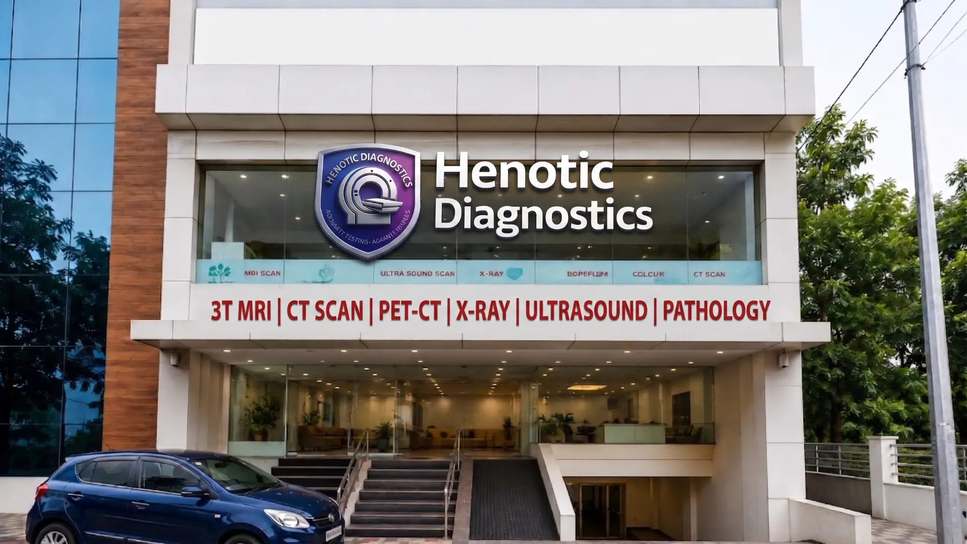
patient Lounge & Executive Reception for Diagnostic Studies Center
Climate-controlled reception area with wheelchair access and instant registration desks.
what Is Diagnostic Imaging?
definition of Medical Imaging
Imaging examinations encompass a set of non-invasive procedures that capture structural and functional representations of the human body. These techniques allow physicians to diagnose medical conditions, evaluate traumatic injuries, and monitor response to therapeutic interventions without surgical exploration.
how Diagnostic Radiological Examinations Works
As a result, because of this, different modalities utilize distinct physical principles to form anatomical images. X-ray and CT scans pass controlled energy beams through tissues of varying densities, producing contrast between bone, soft tissue, and air. In contrast, Magnetic Resonance Imaging utilizes radio waves within a high-strength magnetic field to align hydrogen nuclei in cellular fluids. At the same time, ultrasound relies on high-frequency sound waves reflecting off internal tissue boundaries.
what Medical Imaging Scans Can Evaluate
At the same time, in clinical practice, radiology studies support diagnostic evaluation across virtually all medical specialties.
- Structural integrity of bones, joints, and spinal columns
- Soft-tissue lesions, muscle tears, and ligamentous injuries
- Neurological conditions involving brain parenchyma and peripheral nerves
- Vascular blood flow, arterial narrowing, and venous thrombosis
- Abdominal organ morphology including liver, kidneys, and pancreas
- Cardiovascular anatomy, coronary patency, and cardiac valve motion
imaging vs. Overall, diagnostic Testing — Diagnostic Scanning Services
Although laboratory blood tests measure chemical bio-markers, imaging studies provide spatial visualization of anatomical structures. In particular, combining clinical laboratory analysis with modern advanced imaging yields a far more accurate diagnostic picture for treating physicians.
types of Medical Imaging Modalities
Therefore, because medical technology has evolved rapidly, multiple specialized imaging modalities are currently available. Depending on the patient’s symptoms and clinical indications, physicians select the modality that provides the clearest diagnostic answers.
Magnetic Resonance Imaging (MRI Diagnostic Scans)
Accordingly, mRI provides detailed soft-tissue contrast without ionizing radiation. High-field 3T MRI systems offer enhanced spatial resolution for complex neurological imaging, joint cartilage assessment, and pelvic disease staging. Furthermore, advanced functional sequences including Diffusion Tensor Imaging (DTI tractography), MR Spectroscopy (MRS Choline/NAA ratios), and MR Elastography (MRE) deliver quantitative microstructural tissue analysis.
Computed Tomography (CT Diagnostic Studies Scan)
In addition, computed Tomography creates detailed cross-sectional slices of internal organ structures using computer-assisted X-ray processing. Multi-detector 128-slice CT scanners acquire volumetric data in seconds, making CT the preferred modality for emergency trauma screening, acute chest pain, and pulmonary angiography. Notably, Deep Learning Image Reconstruction (DLIR) and ASiR-V™ algorithms eliminate image noise while optimizing iodine attenuation.
Ultrasound Diagnostic Imaging & Sonography
Diagnostic ultrasound uses non-ionizing sound waves and is widely used for fetal and pregnancy imaging when clinically indicated. High-resolution sonography allows real-time visualization of abdominal organs, thyroid nodules, breast tissues, and superficial soft-tissue structures without thermal tissue heating or ionizing radiation.
Color Doppler Medical Imaging Vascular Imaging
Furthermore, color Doppler ultrasound overlays directional blood flow velocity onto structural sonograms. Vascular specialists use Doppler studies to detect deep vein thrombosis (DVT), carotid artery stenosis, peripheral arterial occlusion, and placental vascular resistance.
Digital Radiography (X-Ray Imaging Examinations)
Digital X-ray remains the foundational imaging test for primary bone fracture assessment, joint arthritis evaluation, and pulmonary infection screening. Modern flat-panel digital detectors significantly reduce exposure while producing instant digital images for immediate radiologist review.
Mammography Diagnostic Scans & Advanced Breast Imaging
Digital Mammography uses low-energy X-rays tailored specifically for breast tissue evaluation. Moreover, digital breast tomosynthesis (3D mammography) captures multiple thin image slices, improving microcalcification detection in dense breast tissue.
PET-CT Radiology Studies / Nuclear Medicine
Specifically, Positron Emission Tomography combined with CT (PET-CT) merges functional metabolic data with anatomical cross-sections using advanced FDG PET Scan radiotracers. PET-CT scans are instrumental in clinical oncology for tumor staging, quantitative SUVmax metabolic tumor volume (MTV) profiling, and baseline evaluation conforming to RECIST 1.1 and PERCIST 1.0 oncology guidelines.
Cardiac Imaging & Functional Echo
Non-invasive cardiac imaging includes 2D Echocardiography, Tissue Doppler Imaging, and cardiac CT angiography. Cardiologists can precisely measure ejection fraction, assess myocardial wall motion, evaluate valve regurgitation, and non-invasively visualize coronary artery narrowing.
turnaround Time SLAs & Instant Report Badges
45–60 Min STAT SLA for Emergency Cases
Consequently, emergency trauma & acute stroke CT/MRI reports issued rapidly for referring clinicians.
4–6 Hour Routine TAT for Outpatients
As a result, comprehensive outpatient diagnostic reports completed same-day by senior radiologist.
instant DICOM Cloud Link
Therefore, high-resolution diagnostic image web links sent straight to your phone via SMS & WhatsApp.
Radiologist Second Opinion Request & SLA Calculator
Additionally, our Radiologist Second Opinion Request & SLA Calculator provides real-time turnaround time estimates for sub-specialty diagnostic consultations. As a result, referring physicians and patients receive rapid, verified specialist interpretations within guaranteed SLA windows.
your Diagnostic Journey in 4 Simple Steps
In fact, from booking your appointment to receiving your signed digital report, we ensure a transparent, comfortable care experience:
60-Second Online Scan Booking
Notably, book via phone, WhatsApp, or online. For instance, receive instant prep instructions straight to your phone.
rapid Front-Desk Patient Check-In
Dedicated reception desk with zero waiting time and private clinical changing cubicles.
gentle, Low-Dose Scan Protocols
For example, state-of-the-art 3T MRI or 128-slice CT scan conducted with continuous technologist care.
same-Day Digital Report Delivery
Signed PDF report and interactive 3D DICOM cloud web links delivered via SMS & WhatsApp.
what Our Patients Say — Patient Testimonials & Clinical Experiences
Hear how our medical imaging team supports patients through clear communication, efficient scheduling, and precise diagnostic reporting.
Overall, “I underwent an MRI spine scan at Henotic Diagnostics. The team was extremely polite and professional.”
Ultimately, — Rajesh M., Kharghar
“My doctor requested an abdominal ultrasound and Doppler study. Hence, i received the digital report on WhatsApp within hours.”
In fact, because of this, — Sunita S., Navi Mumbai
“The CT scan procedure was fast and well-coordinated. At the same time, highly recommend Henotic Diagnostics.”
In particular, — Amit K., Panvel
frequently Asked Questions About Advanced Imaging
Here are answers to common questions regarding diagnostic imaging scans, safety considerations, preparation, and reporting procedures.
what is medical imaging?
Accordingly, diagnostic imaging refers to non-invasive techniques used to create visual representations of internal body structures. These techniques include MRI, CT, ultrasound, X-ray, and PET-CT, allowing clinicians to evaluate organs, bones, and tissues.
what are the main types of medical imaging?
The primary modalities include Magnetic Resonance Imaging (MRI), Computed Tomography (CT), Diagnostic Ultrasound, Digital Radiography (X-Ray), Mammography, Color Doppler, PET-CT, and Echocardiography.
does MRI Use Ionizing Radiation?
No. MRI operates using strong magnetic fields and radiofrequency pulses. In addition, it does not expose patients to ionizing radiation.
What is the Difference Between MRI and CT Scans?
Furthermore, mRI provides detailed soft-tissue contrast without ionizing radiation. CT uses X-rays to acquire fast cross-sectional images, making CT preferred for acute trauma, bone fractures, and chest conditions.
is Diagnostic Ultrasound Safe During Pregnancy?
Overall, yes. Diagnostic ultrasound uses non-ionizing sound waves and is widely used for fetal and pregnancy imaging when clinically indicated.
does CT Scan Use Ionizing Radiation?
Hence, yes. Moreover, cT scans utilize ionizing X-rays. Henotic Diagnostics employs low-dose scan protocols to minimize radiation exposure while maintaining high diagnostic clarity.
when is Contrast Media Required for Diagnostic Scans?
Specifically, contrast agents are administered when doctors need to highlight vascular structures, tissue perfusion, or tumor boundaries. Consequently, renal function tests are performed prior to contrast administration for patient safety.
how to Prepare for Your Diagnostic Scan
As a result, preparation depends on the modality. MRI requires removing metallic items, CT contrast scans may require fasting, and abdominal ultrasounds usually require 6 hours of fasting.
who Interprets Diagnostic Radiology Scans?
Therefore, medical imaging scans are interpreted by qualified radiologists who prepare diagnostic reports for referring clinicians.
what Does ‘Impression’ Mean in a Radiology Report?
The ‘Impression’ section provides the radiologist’s overall diagnostic conclusion summarizing the significant findings observed across the examination images.
henotic Diagnostic Center & Corporate Imaging Examinations Booking Office
Visit our state-of-the-art diagnostic imaging facility in Sector 15, Kharghar for 3T MRI, 128-Slice CT, 3D Ultrasound, and PET-CT scans with zero waiting time.
daily Medical Physics Quality Assurance & ACR Phantom Testing
In addition, our medical physics team conducts daily ACR phantom QA calibrations, verifying Signal-to-Noise Ratios (SNR) for 3T MRI systems and line-pair spatial resolution (lp/mm) for 3D mammography detectors.
our Diagnostic Scans Accreditations & Quality Standards
Radiology Quality and Safety standards at Henotic Diagnostics strictly align with national statutory regulatory authorities and international quality frameworks.
recognized Statutory Bodies & Quality Accreditation Authorities
.webp)
aERB Compliant
In fact, radiation Safety & Diagnostic Equipment Quality Assurance

nABL ISO 15189
Notably, laboratory Quality & Diagnostic Competency Accreditation

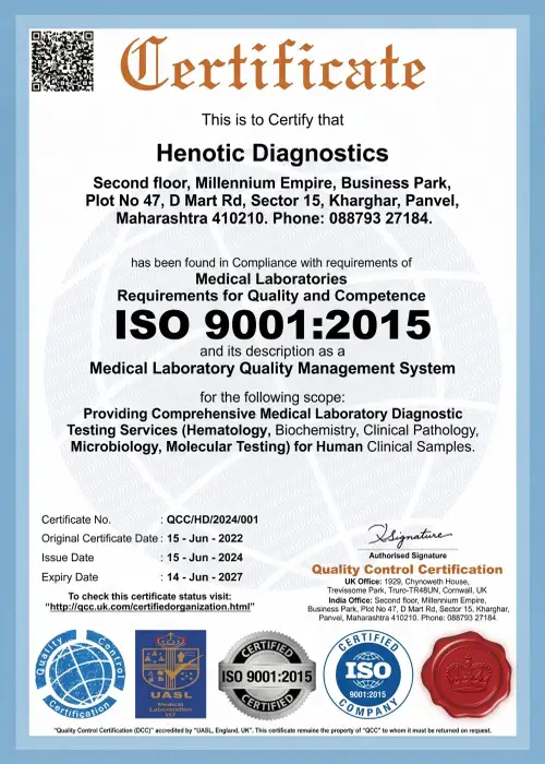
iSO 9001:2015
For instance, international Quality Management System Certified

iSUOG Member
For example, international Society of Ultrasound in Obstetrics & Gynecology

nABH Compliant
Overall, healthcare Facility Quality & Patient Safety Protocols

pCPNDT Registered
Ultimately, full Compliance with Pre-Conception & Pre-Natal Diagnostic Techniques Act
official Quality & Safety Certification Documentation



diagnostic & Radiological Examinations Center Services
Learn About our complete range of diagnostic imaging services and specialized scans at Henotic Diagnostics. Furthermore, bone mineral densitometry offers quantitative FRAX® 10-Year Fracture Risk calculation, Vertebral Fracture Assessment (VFA), Trabecular Bone Score (TBS), Femoral Neck T-Score measurement, and strict ISCD Reporting Standards:
- As a result, → Magnetic Resonance Imaging (3T MRI) Services
- Therefore, → 128-Slice Computed Tomography (CT Scan)
- In fact, → 3D/4D Diagnostic Ultrasound & Fetal Sonography
- Notably, → Color Doppler Vascular Flow Studies
- For instance, → Digital Radiography & Contrast X-Ray Examinations
- For example, → Digital 3D Breast Mammography Screening
- Overall, → PET-CT Whole Body Oncology Scanning
- Ultimately, → 2D Echocardiography Services
- Hence, → CT Coronary Angiography Services