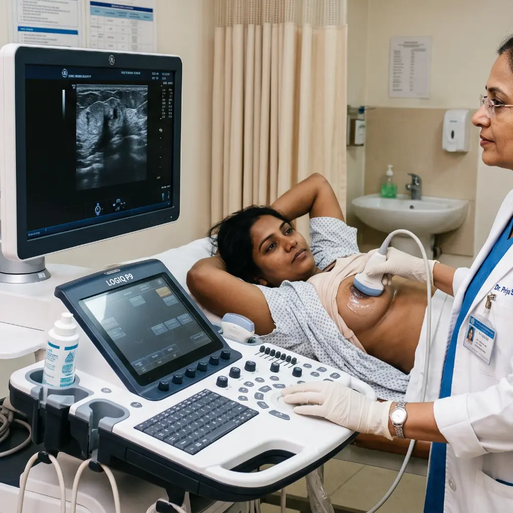
Advanced Bilateral Sonomammography
Accurate, radiation-free, high-resolution breast ultrasound for detailed evaluation of breast tissue, lumps, cysts and other clinically relevant findings, with interpretation by qualified radiology professionals.
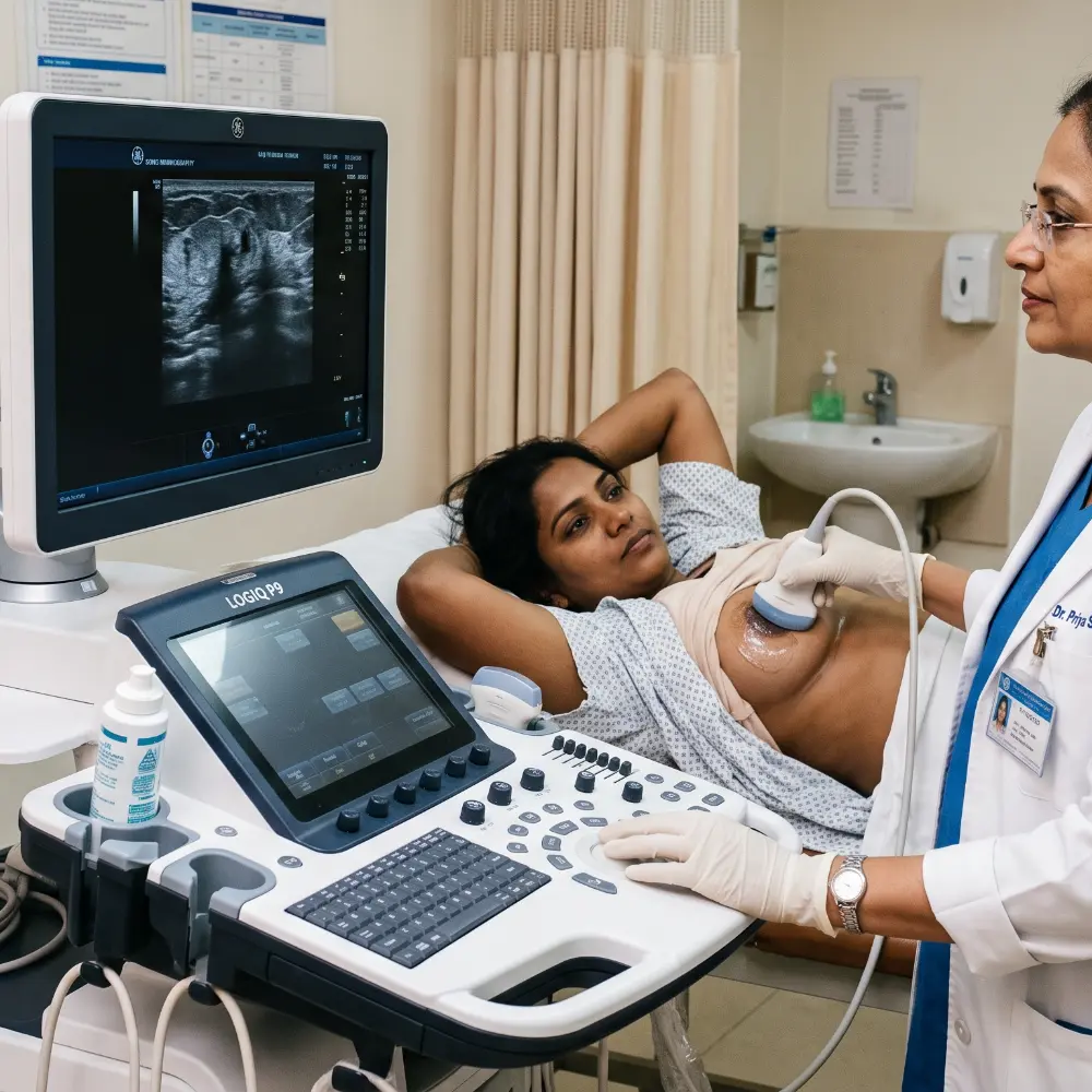
Advanced Breast Ultrasound for Detailed Breast Evaluation
Sono Mammography, also known as breast ultrasound, is an ultrasound-based examination used to evaluate breast tissue, breast lumps, cysts, masses and other breast abnormalities.
Radiation-Free Imaging
Breast ultrasound uses high-frequency sound waves and does not use ionizing radiation.
Targeted Evaluation
Ultrasound can provide detailed assessment of clinically relevant breast findings and focal abnormalities.
Professional Reporting
Acquired images are assessed and interpreted by qualified radiology professionals.
Sono Mammography at a Glance
| Service Information | Details |
|---|---|
| Examination | Sono Mammography / Breast Ultrasound |
| Imaging method | Ultrasound |
| Ionizing radiation | No |
| Fasting | Not required for routine examination |
| Preparation | Minimal |
| Typical duration | Approximately 15–30 minutes |
| Interpretation | Radiologist reporting |
| Digital reports | Available |
| WhatsApp / Email delivery | Available |
| Centre availability | Open 24 hours |
| Appointment | Prior booking recommended |
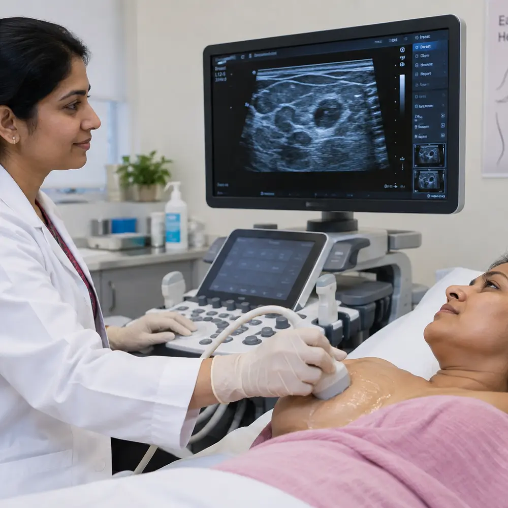
What Can Sono Mammography Evaluate?
Breast ultrasound can help evaluate a range of breast findings. The appropriate examination depends on the patient’s age, symptoms, medical history, risk factors and clinical question.
- Breast lumps
- Breast cysts
- Solid breast masses
- Localised breast pain
- Palpable breast abnormalities
- Focal breast abnormalities
- Previously identified breast lesions
- Selected mammography findings requiring further evaluation
- Selected findings in patients with dense breast tissue
Why Is Sono Mammography Performed?
Sono Mammography may be recommended when a clinician needs additional information about a breast symptom, palpable finding or imaging abnormality.
New Breast Lump
Ultrasound may help characterise a palpable or clinically relevant breast finding.
Breast Cyst
Ultrasound can help assess whether a finding has a cystic or solid appearance.
Focal Breast Pain
Targeted ultrasound may be used when breast symptoms require imaging evaluation.
Abnormal Mammography
Ultrasound may provide additional information about selected mammographic findings.
Dense Breast Tissue
Ultrasound may be used as an additional examination in selected patients when clinically appropriate.
Follow-Up
Ultrasound can be used to reassess selected previously identified breast lesions.
Sono Mammography and Mammography Are Different Examinations
Sono Mammography
- Uses ultrasound waves
- Does not use ionizing radiation
- Produces real-time ultrasound images
- Useful for targeted evaluation
- Can help distinguish cystic and solid appearances
Mammography
- Uses low-dose X-rays
- Uses ionizing radiation
- Produces X-ray images of breast tissue
- Important breast imaging modality
- Used for screening and diagnostic evaluation when appropriate
Breast Cysts and Solid Lesions
Ultrasound can help the radiologist assess the imaging characteristics of a breast finding.
| Ultrasound Assessment | Clinical Information |
|---|---|
| Cystic appearance | Helps identify fluid-containing lesions |
| Solid appearance | Allows assessment of morphology and other characteristics |
| Lesion margins | Contributes to imaging characterisation |
| Shape | Helps describe imaging appearance |
| Internal features | Provides additional diagnostic information |
| Size | Allows measurement and documentation |
| Location | Identifies the anatomical site |
Sono Mammography for Dense Breasts
Breast density can affect the appearance of breast tissue on mammography. Ultrasound may be used as an additional imaging examination in selected patients when clinically appropriate.
The decision to perform additional breast imaging should be based on the patient’s clinical circumstances and healthcare professional’s recommendation.
Book Breast Ultrasound
Sono Mammography During Pregnancy
Ultrasound does not use ionizing radiation and may be used when breast imaging is clinically required during pregnancy.
If you are pregnant or think you may be pregnant, inform the radiology team before the examination.
The appropriate imaging pathway depends on the patient’s symptoms and clinical circumstances.
How Is Sono Mammography Performed?
The examination is generally straightforward and does not require fasting.
Registration
Appointment and patient details are verified.
Clinical History
Relevant symptoms and previous imaging may be reviewed.
Positioning
You are positioned comfortably for the examination.
Ultrasound Scan
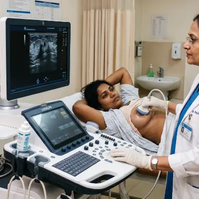
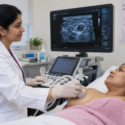
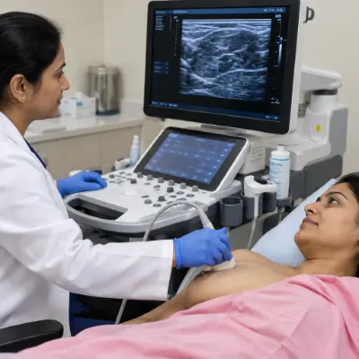
Gel is applied and the transducer is moved across the breast.
Reporting
Images are assessed and findings documented in the report.
What Happens During the Examination?
| Examination Stage | What You Can Expect |
|---|---|
| Registration | Patient and appointment details are verified |
| Clinical history | Relevant symptoms and previous imaging may be discussed |
| Positioning | Comfortable examination position |
| Gel application | Ultrasound gel is applied to the skin |
| Scanning | Transducer is moved across the breast |
| Radiologist review | Images are assessed for relevant findings |
| Reporting | Findings are documented in the report |
Sono Mammography Preparation
Routine Sono Mammography generally requires minimal preparation.
Fasting
Fasting is not required for routine Sono Mammography.
Food & Water
Normal food intake and hydration are generally permitted.
Previous Reports
Bring previous mammography or ultrasound reports when available.
Previous Images
Bring previous imaging images or CDs where available.
Prescription
Bring your doctor’s prescription or referral if available.
Pregnancy
Inform the radiology team if you are pregnant or possibly pregnant.
Professional Reporting & Digital Report Delivery
Following image acquisition, the examination is assessed by the reporting radiologist and the findings are documented in the diagnostic report.
- Radiologist interpretation
- Digital report
- WhatsApp report delivery
- Email report delivery
- Patient portal where available
- Hardcopy collection at centre on request
Reporting turnaround: Examination-specific reporting time can depend on the appointment, examination findings and radiologist workflow.
Understanding Your Report
A Sono Mammography report may contain the clinical indication, breast tissue appearance, lesion location, measurements, shape, margins, internal characteristics, radiologist impression and recommendations where indicated.
Any sample report published online should be completely anonymised and contain no personally identifiable patient information.
Our Reporting Radiologists
Breast ultrasound findings are interpreted in the context of the clinical indication and available previous imaging.

Dr Pratibha Patil
16+ years experience; specialist focus in Women’s Imaging & BI-RADS.

Dr Amulya Patil
11+ years experience; breast ultrasound and USG-guided FNAC clinical focus.
Accreditations & Certifications
Display certification and accreditation logos only when the corresponding credential is current and applicable.
 NABL
NABL
 CAP
CAP
 ISO 9001:2015
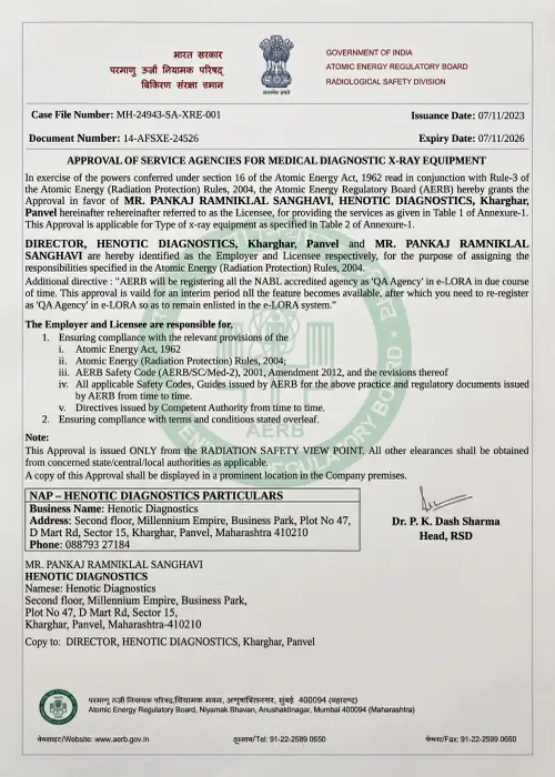 AERB
AERB
 ISUOG
ISUOG
 PCPNDT
Sonomammography Imaging
Why Choose Henotic Diagnostics?
Qualified Radiology Professionals
Professional radiology interpretation for the examination.
Modern Ultrasound Imaging
Breast ultrasound performed using diagnostic ultrasound imaging technology.
Patient Privacy
Breast imaging is performed with attention to privacy, dignity and professional patient handling.
Digital Reporting
Digital report delivery options including WhatsApp and email where available.
Previous Image Comparison
Previous reports and images can be reviewed when provided and clinically appropriate.
24-Hour Centre
Henotic Diagnostics is open 24 hours according to the supplied centre information.
Your Sono Mammography Patient Journey
Book
Call or WhatsApp the centre.
Confirm
Receive appointment details.
Visit
Arrive at the diagnostic centre.
Scan
Complete the breast ultrasound examination.
Report
Receive the diagnostic report through available channels.
When Further Evaluation May Be Required
Sono Mammography is an imaging examination and does not establish a final diagnosis for every breast abnormality.
| Possible Next Step | Purpose |
|---|---|
| Clinical examination | Correlate imaging with symptoms and examination findings |
| Mammography | Additional breast imaging when clinically appropriate |
| Additional ultrasound | Further targeted evaluation |
| MRI | Further evaluation when clinically indicated |
| Follow-up imaging | Monitor selected findings over time |
| Specialist consultation | Clinical assessment and management |
| Biopsy | Tissue diagnosis when indicated |
Sono Mammography — Direct Answers
What is Sono Mammography?
Sono Mammography is an ultrasound examination of the breast that uses high-frequency sound waves to produce images of breast tissue and evaluate findings such as lumps, cysts and masses.
Is fasting required?
No. Fasting is not required for routine Sono Mammography.
Does Sono Mammography use radiation?
No. Breast ultrasound uses sound waves and does not use ionizing radiation.
How long does the examination take?
A typical examination takes approximately 15–30 minutes, although the duration can vary.
Is Sono Mammography painful?
It is generally well tolerated. Some patients may experience mild pressure while the transducer is moved over the breast.
What can breast ultrasound evaluate?
It can help evaluate breast lumps, cysts, solid masses, focal abnormalities and other clinically relevant breast findings.
Is Sono Mammography the same as mammography?
No. Sono Mammography uses ultrasound, while mammography uses X-rays. They have different clinical roles and may sometimes be used together.
Can breast ultrasound be performed during pregnancy?
Ultrasound does not use ionizing radiation and may be used when breast imaging is clinically indicated during pregnancy. The appropriate examination should be determined by a healthcare professional.
Real Patient Experiences
“[INSERT VERIFIED PATIENT TESTIMONIAL]”
— [PATIENT NAME / INITIAL AS PER CONSENT]“[INSERT VERIFIED PATIENT TESTIMONIAL]”
— [PATIENT NAME / INITIAL AS PER CONSENT]“[INSERT VERIFIED PATIENT TESTIMONIAL]”
— [PATIENT NAME / INITIAL AS PER CONSENT]Need a Breast Ultrasound?
Contact Henotic Diagnostics to enquire about Advanced Bilateral Sonomammography.
Diagnostic Centre Location
Henotic Diagnostics
Address
Second Floor, Millennium Empire, Business Park,
Plot No. 47, D Mart Road, Sector 15,
Kharghar, Panvel, Maharashtra 410210
Phone
08879327184
Opening Hours
Open 24 Hours
This page provides general educational information about Sono Mammography and breast ultrasound. It is not a substitute for medical consultation, diagnosis or treatment.
The appropriate breast-imaging examination depends on the patient’s symptoms, age, medical history, risk factors and clinical circumstances.
Imaging findings should be interpreted by an appropriately qualified healthcare professional. Additional investigations may be recommended when clinically indicated.