First of all, an MRI Foot scan provides a non-invasive, high-resolution diagnostic evaluation of forefoot, midfoot, and hindfoot soft tissue structures, tendons, and small tarsal bones. Essentially, this advanced 3T foot MRI scan offers superior musculoskeletal detail compared to standard X-rays. As a result, radiologists accurately differentiate severe plantar fasciitis, Morton’s neuroma, Lisfranc ligament tears, occult metatarsal stress fractures, and diabetic foot osteomyelitis.
Furthermore, you can book your Magnetic Resonance Imaging of the Foot scan at Henotic Diagnostics for complete clinical precision and patient comfort. In addition, our accredited diagnostic facility operates 24/7 to provide prompt, seamless appointment booking.
Overview of MRI Foot Scanning for Foot & Ankle Health
To begin with, an MRI Foot scan delivers vital diagnostic insights when evaluating complex podiatric and orthopedic conditions. At the same time, specialists at Henotic Diagnostics utilize multi-channel 3T magnetic resonance scanners to generate crisp multiplanar cross-sectional images of the plantar fascia, intermetatarsal nerves, tarsal joints, and intrinsic foot musculature.
What Does an MRI Foot Scan Evaluate?
Specifically, the scan evaluates plantar fascial thickening, calcaneal spurs, Morton’s intermetatarsal neuromas, Lisfranc joint complex disruption, tarsal tunnel nerve compression, tendon tears (Achilles, tibialis anterior/posterior, peroneus), and diabetic soft tissue ulcers extending to bone (osteomyelitis). Consequently, orthopedic surgeons, podiatrists, and sports medicine physicians rely on high-resolution T1, T2, STIR, and PD Fat-Sat images to plan effective non-surgical or operative care. Furthermore, multiplanar MR images distinguish bone marrow edema from true cortical fractures. In fact, to review musculoskeletal radiology guidelines, consult the RadiologyInfo resource.
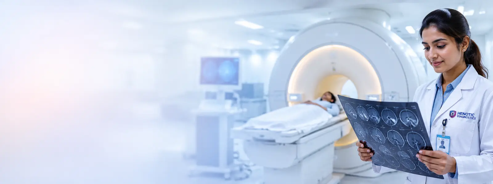
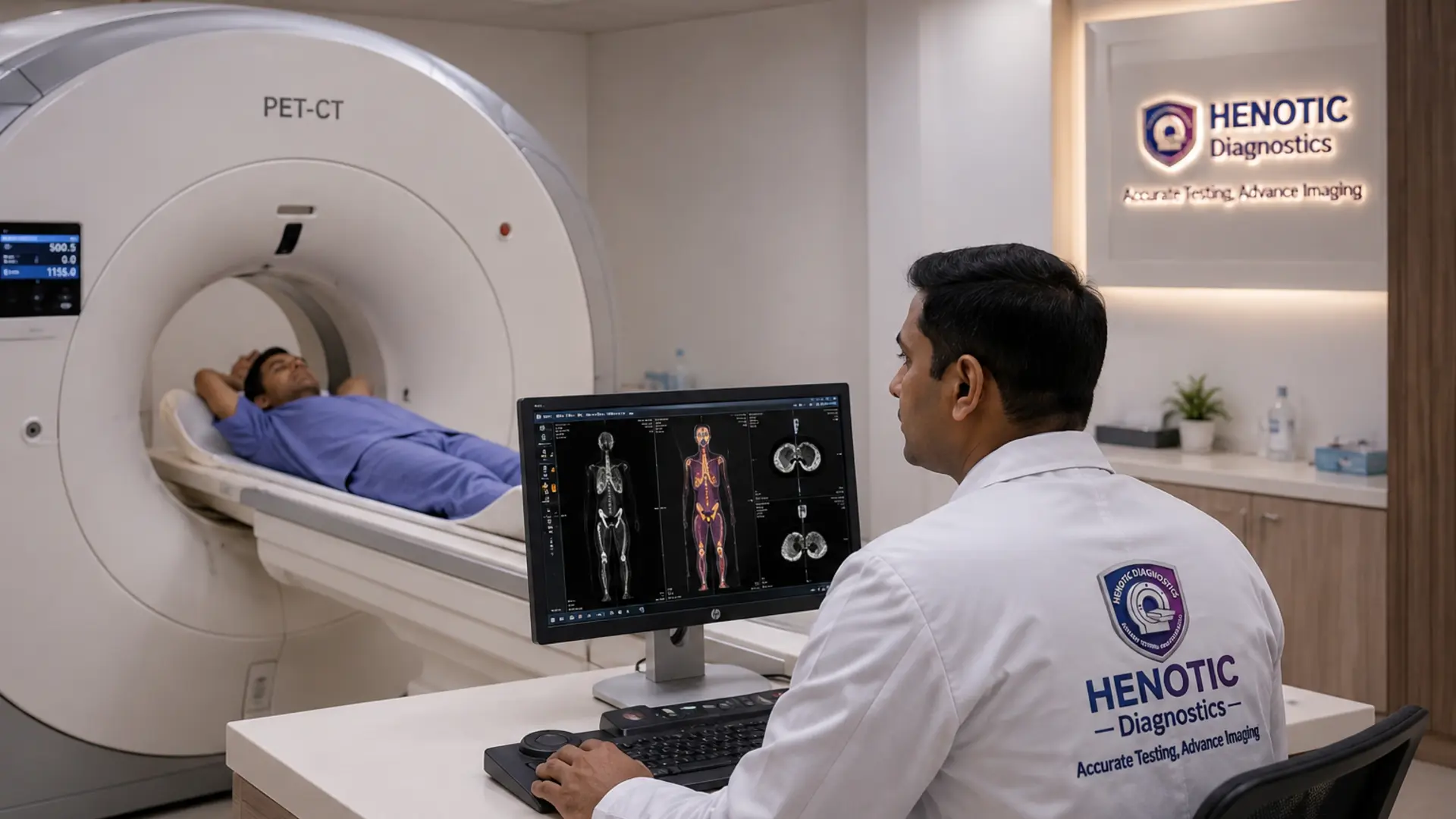
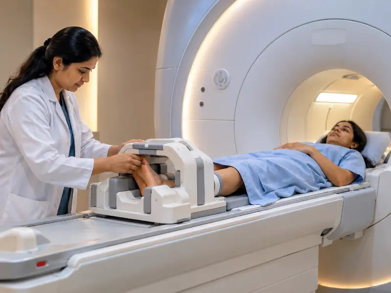
Furthermore, private 3T MRI scanning suite ensures patient comfort and high diagnostic precision.
What is an MRI Foot Scan?
In general, an Magnetic Resonance Imaging of the Foot scan is a non-invasive diagnostic procedure that captures multiplanar magnetic resonance cross-sections of the entire foot architecture. Essentially, radiofrequency pulses align hydrogen protons in tendons, ligaments, cartilage, and bone marrow, producing exceptionally detailed images of the Lisfranc joint, Chopart joint, and metatarsophalangeal (MTP) joints. Therefore, physicians identify subtle soft tissue injuries that plain X-rays miss. Additionally, this scan involves zero ionizing radiation.
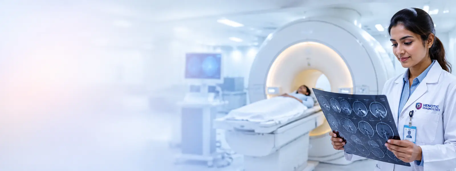
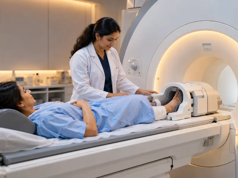
Additionally, wide-bore 3T MRI scanners deliver exceptional signal resolution and patient comfort.
Key Diagnostic Parameters & Findings in MRI Foot Scanning
Overall, radiologists analyze four key structural and pathological parameters on multiplanar 3T MRI Foot sequences:
MRI Foot Key Clinical Indicators Examined
✓
Plantar Fascia & Calcaneal Insertion: First, high PD Fat-Sat signal and fascial thickening (>4mm) confirm severe chronic plantar fasciitis or rupture.
✓
Morton’s Intermetatarsal Neuroma: Second, teardrop-shaped soft tissue masses in the 3rd or 2nd web space confirm intermetatarsal plantar nerve entrapment.
✓
Lisfranc & Midfoot Ligament Disruption: In addition, coronal and axial views evaluate midfoot instability, cuneiform subluxation, and Lisfranc ligament tears.
✓
Metatarsal Stress Fractures & Osteomyelitis: Finally, high T2 STIR marrow edema confirms early stress fractures or diabetic bone infection beneath skin ulcers.
In addition, real-time multiplanar 3T MRI scans accurately map foot soft tissue and tarsal bone structures.
When Is an MRI Foot Scan Recommended?
For example, podiatrists and orthopedic specialists order an MRI Foot scan when patients experience chronic heel pain unresponsive to therapy, sharp forefoot burning pain (suspected Morton’s neuroma), midfoot swelling following trauma (suspected Lisfranc injury), or persistent metatarsal pain in runners. In particular, doctors request testing prior to foot surgery, or when evaluating diabetic foot ulcers for underlying bone osteomyelitis or deep abscess formation.
Moreover, high-resolution 3T MRI Foot scanning is recommended for complex tendon, ligament, and osteomyelitis evaluations.
Step-by-Step Patient Preparation and Prerequisites
First of all, proper preparation ensures a smooth MRI Foot examination. Prior to entering the scanner room, patients complete a safety screening checklist to identify any metallic implants, pacemakers, or metallic foreign bodies.
✓
Remove Metal Objects: First, remove footwear, ankle bracelets, body piercings, hairpins, watches, and metallic clothing fasteners.
✓
Inform Surgical Hardware: Second, inform our technologist if you have orthopedic screws, plates, or joint pins in your foot or ankle.
✓
Claustrophobia Support: In addition, report any anxiety in enclosed spaces so our staff can position your upper body outside the wide-bore scanner.
Fasting and Contrast Injection Conditions
In general, routine non-contrast MRI Foot scans for tendon, ligament, or stress fracture evaluation require no fasting whatsoever. However, if your doctor orders a contrast-enhanced MRI (CE-MRI Foot) to evaluate diabetic foot osteomyelitis, soft tissue abscesses, or foot tumors, specific clinical conditions apply:
Contrast Gadolinium Safety Protocol:
First, maintain 4 hours of strict fasting (water permitted) prior to contrast administration.
Second, provide recent Serum Creatinine / eGFR lab results to confirm normal kidney clearance.
In addition, a small IV cannula is placed safely into an arm vein for gadolinium contrast injection during scanning.
Furthermore, contrast gadolinium sharply delineates soft tissue abscess walls and bone marrow infection.
What to Expect During the Procedure
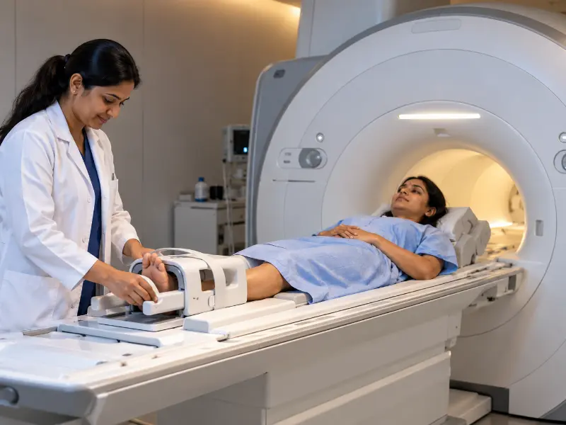
Overall, the MRI Foot scan is painless, comfortable, and zero-radiation. You will lie comfortably on your back on the padded scanner table, with your symptomatic foot placed gently into a dedicated extremity coil. Because foot imaging allows feet-first positioning, your head remains near the open end of the scanner. Noise-reducing headphones are provided to buffer magnet tapping sounds. The scan takes 25 to 40 minutes, during which you remain still. Additionally, an emergency squeezable bulb is held in your hand at all times.
Advanced Medical Technology: Machines and Models
At Henotic Diagnostics, we utilize state-of-the-art 3 Tesla (3T) wide-bore MRI systems engineered by Siemens and GE Healthcare. Specifically, our machines feature high-density dedicated foot/ankle RF coils and specialized pulse sequences (T1, T2, STIR, and Proton Density Fat-Sat). Therefore, our scanners resolve sub-millimeter plantar fascia fibers and detect subtle intermetatarsal neuromas that low-field scanners miss.
Post-Procedure Care & Aftercare Guidelines
Fortunately, recovery after an MRI Foot scan is immediate. Once the scan is complete, you may resume normal activities, return to work, and drive home without downtime.
✓
Resume Normal Routine: First, you can immediately resume your daily meals and activities.
✓
Flush Contrast Fluid: Second, if contrast injection was administered, drink 2 to 3 liters of water over the next 24 hours to eliminate contrast fluid cleanly through your kidneys.
✓
IV Site Care: In addition, keep the small adhesive bandage on your arm IV site for 2 to 3 hours to prevent minor bruising.
Diagnostic Reporting and Hardcopy Availability
In addition, our digital workflow delivers fast, high-precision reporting. Following your scan, complete DICOM image sets are uploaded to our PACS server. Chief Radiologist Dr. Pratibha Patil evaluates axial, coronal, and sagittal cross-sections, delivering detailed diagnostic reports within 2 to 4 hours. Furthermore, printed laser hardcopy films and digital USB/cloud links are available at reception.
Second Opinion & Subspecialty Consultations
Besides routine reporting, complex Lisfranc injuries, diabetic osteomyelitis, or surgical planning require expert subspecialty review. At Henotic Diagnostics, we provide dedicated second opinions and direct peer-to-peer case discussions with treating podiatrists and orthopedic surgeons:
Subspecialty Musculoskeletal Services:
First, expert MSK radiologist second opinion reviews on external foot MRI films or DICOM data.
Second, direct doctor-to-doctor consultations between our radiologist and your attending orthopedic surgeon.
In addition, multiplanar mapping of Lisfranc ligament tears prior to surgical reconstruction.
Furthermore, instant encrypted digital image sharing for emergency clinical reviews.
Why Choose Us for Your Care?
Choosing Henotic Diagnostics for your MRI Foot scan guarantees top-tier imaging technology and compassionate patient care:
✓
5.0 Star Rated Patient Care: First, over 10,000 satisfied patients rate us top for cleanliness, efficiency, and care.
✓
Subspecialty Musculoskeletal Radiology: Second, reports are interpreted by expert radiologists specializing in foot and ankle imaging.
✓
NABL & AERB Accredited Safety: In addition, our facility strictly meets international diagnostic and radiation-free standards.
✓
24/7 Operations & Rapid TAT: Finally, 24/7 slot booking ensures rapid scan completion and 2-hour report turnaround.
Transportation & Center Accessibility
Moreover, our diagnostic facility in Kharghar, Navi Mumbai, is designed for smooth accessibility:
✓
Prime Location & Landmark: First, located on 2nd Floor, Millennium Empire, Business Park, D Mart Road, Sector 15, Kharghar.
✓
Dedicated Parking: Second, spacious ground and basement parking areas are reserved for patient vehicles.
✓
Full Wheelchair Accessibility: In addition, wide elevators, barrier-free ramps, and staff assistance ensure complete wheelchair mobility.
✓
24/7 Emergency Scheduling: Finally, our MRI department runs continuously to accommodate urgent STAT foot scans.
Emergency Warning Symptoms for Foot & Ankle Health
First, When to Consult Your Podiatrist Urgently
Ideally, you should seek urgent medical evaluation if you experience inability to bear weight after midfoot trauma, a popping sound with sudden heel loss of function (suspected Achilles tendon rupture), expanding redness/warmth from a diabetic foot ulcer, or severe deep pain suggesting acute osteomyelitis. In addition, immediate 3T MRI Foot scanning is vital to prevent long-term disability.
MRI Foot Test Cost Guide
First, Diagnostic Foot Service
Second, Approximate Cost (₹)
First, Standard Non-Contrast 3T MRI Foot Scan
First, standard MRI Foot scan cost is ₹6,500 – ₹8,500.
Finally, expert radiologist consultation fee is ₹1,000.
Comparison: Foot X-Ray vs. MRI Foot
On one hand, X-ray Foot excels at demonstrating major displaced bone fractures and calcaneal bone spurs. On the other hand, an MRI Foot scan provides superior soft tissue characterization, easily revealing plantar fascia tears, Morton’s neuroma, early bone marrow edema, and Lisfranc ligament injuries without radiation.
First, Feature
Second, Foot X-Ray
Third, MRI Foot (3T MSK MRI)
First, Primary Strength
First, X-ray excels at gross bony alignment & spurs.
However, MRI excels at tendons, nerves, & marrow edema.
Second, Early Stress Fracture Detection
Second, X-ray is insensitive to early non-displaced stress lines.
In contrast, MRI detects early stress marrow edema immediately.
In addition, Soft Tissue & Nerve Visualization
In addition, X-ray cannot see plantar fascia or Morton’s neuroma.
Second, MBBS, MD (Radiodiagnosis) · 15+ Years Clinical Experience
Furthermore, your MRI Foot scans and multiplanar reconstructions are analyzed by senior musculoskeletal radiodiagnostic experts. Consequently, comprehensive reports provide podiatrists and orthopedic surgeons with precise anatomical data to evaluate plantar fasciitis, Lisfranc tears, and osteomyelitis.
Our Accreditations and Quality Standards
In addition, certified excellence ensures safety, hygiene compliance, and accurate diagnostic results.
What Our Patients Say
Furthermore, read real feedback from patients who completed MRI Foot scanning with us.
First, ⭐⭐⭐⭐⭐
“First of all, Dr. Pratibha Patil’s team delivered a precise 3T MRI Foot report identifying a Morton’s neuroma that was missed on ultrasound. Very clear findings and quick delivery!”
– Sanjay T.
First, Verified Patient Review
Second, ⭐⭐⭐⭐⭐
“In addition, the feet-first MRI positioning was extremely comfortable. As a runner with a metatarsal stress fracture, the scan was quick and totally painless.”
– Deepika M.
Second, Verified Patient Review
Finally, ⭐⭐⭐⭐⭐
“Furthermore, prompt report delivery within 2 hours. Printed high-resolution laser films and DICOM links were provided right away. Excellent podiatric imaging center.”
– Rahul B.
Finally, Verified Patient Review
Relevant Diagnostic Radiology and Extremity Services
If you require a high precision MRI Foot scan for plantar fasciitis, Morton’s neuroma, Lisfranc injury, or diabetic foot infection, do not delay. In addition, call us today to schedule your appointment.
Henotic Diagnostics
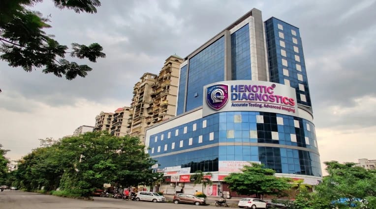
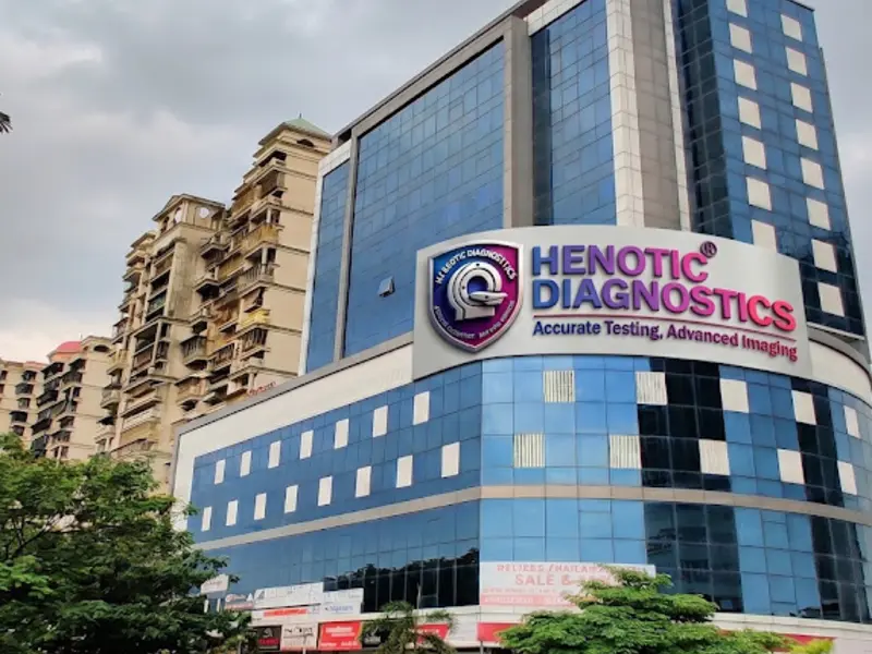
First, Second floor, Millennium Empire, Business Park, Plot No 47, D Mart Rd, Sector 15, Kharghar, Navi Mumbai, Panvel, Maharashtra 410210
You can book your MRI Foot appointment at our Navi Mumbai center through multiple convenient channels: call us directly at 088793 27184, send a WhatsApp message for instant booking, or use the online booking form on our website. Walk-in appointments are also welcome based on slot availability. We recommend advance booking to ensure zero wait times.
Yes, Henotic Diagnostics Navi Mumbai is empanelled with all major insurance companies and TPAs (Third Party Administrators) including Star Health, ICICI Lombard, New India Assurance, HDFC Ergo, Max Bupa, Bajaj Allianz, and many more. We offer cashless processing for MRI Foot where applicable. Please carry your insurance card and a valid ID for seamless processing.
At Henotic Diagnostics Navi Mumbai, most MRI Foot reports are delivered within 2-6 hours for routine tests. Critical and urgent reports are expedited on a priority basis. All reports are delivered digitally via WhatsApp, email, and our secure online patient portal. Hard copies are also available at the center upon request. You will receive an SMS/WhatsApp notification the moment your report is ready.
Henotic Diagnostics Navi Mumbai operates 24 hours a day, 7 days a week, including Sundays and public holidays. This means you can schedule your MRI Foot at a time that is most convenient for you — early morning, late evening, or even overnight for emergency requirements. Our radiology and pathology departments maintain round-the-clock availability.
Yes, Henotic Diagnostics Navi Mumbai is fully accredited by NABL (National Accreditation Board for Testing and Calibration Laboratories) and ISO 9001:2015 certified. Our radiology equipment also complies with AERB (Atomic Energy Regulatory Board) safety standards. These accreditations ensure that every MRI Foot performed at our facility meets the highest national and international quality benchmarks for accuracy, safety, and reliability.
The cost of MRI Foot at Henotic Diagnostics Navi Mumbai starts from just ₹4,500, which is 40-50% lower than the average market price of ₹8,000-12,000. Our pricing is transparent with zero hidden charges. The exact cost may vary depending on the body part being scanned and whether contrast (dye injection) is required. Contact us for an exact quote tailored to your prescription.
For most MRI Foot procedures, no special preparation is needed. However, please note: (1) Remove all metallic objects including jewelry, watches, hairpins, and belts before the scan. (2) Inform your doctor if you have any metallic implants, pacemakers, cochlear implants, or metal clips. (3) For contrast MRI, fasting for 4-6 hours is recommended. (4) Wear comfortable, loose-fitting clothing without metal zippers. (5) Bring your doctor's prescription and any previous scan reports for comparison.
A standard MRI Foot at Henotic Diagnostics Navi Mumbai typically takes 20-45 minutes depending on the body part and clinical indication. A contrast-enhanced MRI may take an additional 10-15 minutes. Our 1.5 Tesla MRI machine ensures high-quality images in optimal scan time. We recommend arriving 15 minutes before your appointment for registration and preparation.
Yes, MRI Foot is completely safe and uses powerful magnetic fields and radio waves — it does NOT use any ionizing radiation (unlike X-ray or CT). This makes MRI one of the safest diagnostic imaging modalities, suitable for repeated use, pregnant women (in certain trimesters with medical guidance), and children. The only contraindication is for patients with certain metallic implants or pacemakers.
We understand that MRI machines can feel confining. At Henotic Diagnostics Navi Mumbai, our radiographers are specially trained to help claustrophobic patients with calming techniques and continuous communication throughout the procedure. In some cases, your referring physician may prescribe a mild sedative before the scan. We also allow a companion to stay in the MRI room for moral support.
A non-contrast (plain) MRI uses magnetic fields alone to produce images. A contrast-enhanced MRI involves injecting a gadolinium-based contrast dye through an IV line, which highlights blood vessels, tumors, and areas of inflammation more clearly. Your doctor will specify whether contrast is needed based on the clinical condition. The contrast dye is generally safe and is excreted by the kidneys within 24 hours. A kidney function test (serum creatinine) may be required before contrast administration.
"My husband was severely claustrophobic and couldn't complete an MRI elsewhere. At Henotic Kharghar, the 70cm wide-bore scanner and calming staff made it completely comfortable. Received reports within 4 hours."
3.0 Tesla Wide-Bore MRI Spine
MK
Meera Krishnan
Vashi, Navi Mumbai
Verified
2:10 min
1 week ago
"Doctor recommended a PET-CT for oncology staging. Henotic team explained the 6-hour fasting and uptake process clearly. The peaceful private uptake room and caring nurses made a stressful day easy."
Digital PET-CT Whole Body
RS
Ravi Sharma
Panvel, Navi Mumbai
Verified
1 week ago
"World-class 3T MRI center in Panvel-Kharghar region. The technologist took time to explain the scan sequences, and the noise-cancellation headphones played relaxing music. Report was verified by Dr. Rajesh Sharma with exceptional clarity."
3.0T MRI Brain with Contrast
AK
Anjali Kulkarni
Kharghar, Sector 15
Verified
2 weeks ago
"Fastest CT scan experience! I arrived with a persistent cough; the staff did the scan in under 3 minutes with ultra-low dose radiation. Had the digital report on WhatsApp before reaching home."
128-Slice HRCT Chest
PJ
Prashant Joshi
Seawoods, Navi Mumbai
Verified
3 weeks ago
"Sports injury while running. The orthopedic surgeon specifically insisted on Henotic's 3.0 Tesla MRI because of their sub-millimeter meniscus cartilage resolution. Extremely satisfied with the diagnostic accuracy."
3.0T MRI Knee Joint (Cartilage Mapping)
DS
Dinesh Singhania
Mumbai
Verified
2 weeks ago
"We traveled from South Mumbai for PET-CT after comparing facilities. The pricing is nearly 40% more transparent than private hospitals, and the machine quality is top-tier Siemens biograph technology. Highly recommended."
18F-FDG PET-CT Whole Body
GM
Gauri Mahajan
Thane West
Verified
1 month ago
"Prompt scheduling, courteous radio-pharmacist, and doctor was available to answer our queries on post-scan radiation precautions. Excellent oncology imaging standard."
DOTA / PSMA PET Scan
SP
Sanjay Patil
Kharghar, Navi Mumbai
Verified
4 days ago
"Phlebotomist arrived right on time at 7:00 AM wearing sterile PPE. Completely painless blood draw for my elderly parents. Digital PDF report delivered by 1:00 PM the same day."
Home Blood Sample Collection (CBC + HbA1c)
PV
Pooja Varma
CBD Belapur
Verified
2 weeks ago
"NABL accredited lab accuracy gives complete peace of mind. Barcode tube labeling was done right in front of me. Clean, automated, and trustworthy."
Thyroid & Vitamin D / B12 Panel
RG
Ramesh & Sunita Gupta
Kamothe, Panvel
Verified
5 days ago
"Booked annual checkups for both my parents. Included 2D Echo, Ultrasound Abdomen, ECG, and full blood work. Completed everything in 2.5 hours with complimentary breakfast. Great hospitality."
Senior Citizen Full Body Health Checkup (85+ Tests)
AB
Amitabh Banerjee
Vashi, Sector 17
Verified
2 weeks ago
"Invaluable early detection! The 2D Echo picked up minor aortic valve thickening which my regular physician corroborated. The doctors at Henotic are true clinicians, not just report printers."