Loading Diagnostics...
Connecting to our secure server
Connecting to our secure server
12 people booked MRI Scan
in Kharghar today • Verified

Access world-class diagnostic technology across our comprehensive network in Mumbai Suburban. Select your nearest neighborhood for priority booking.
Select your specific location to view tailored availability and book your slot.
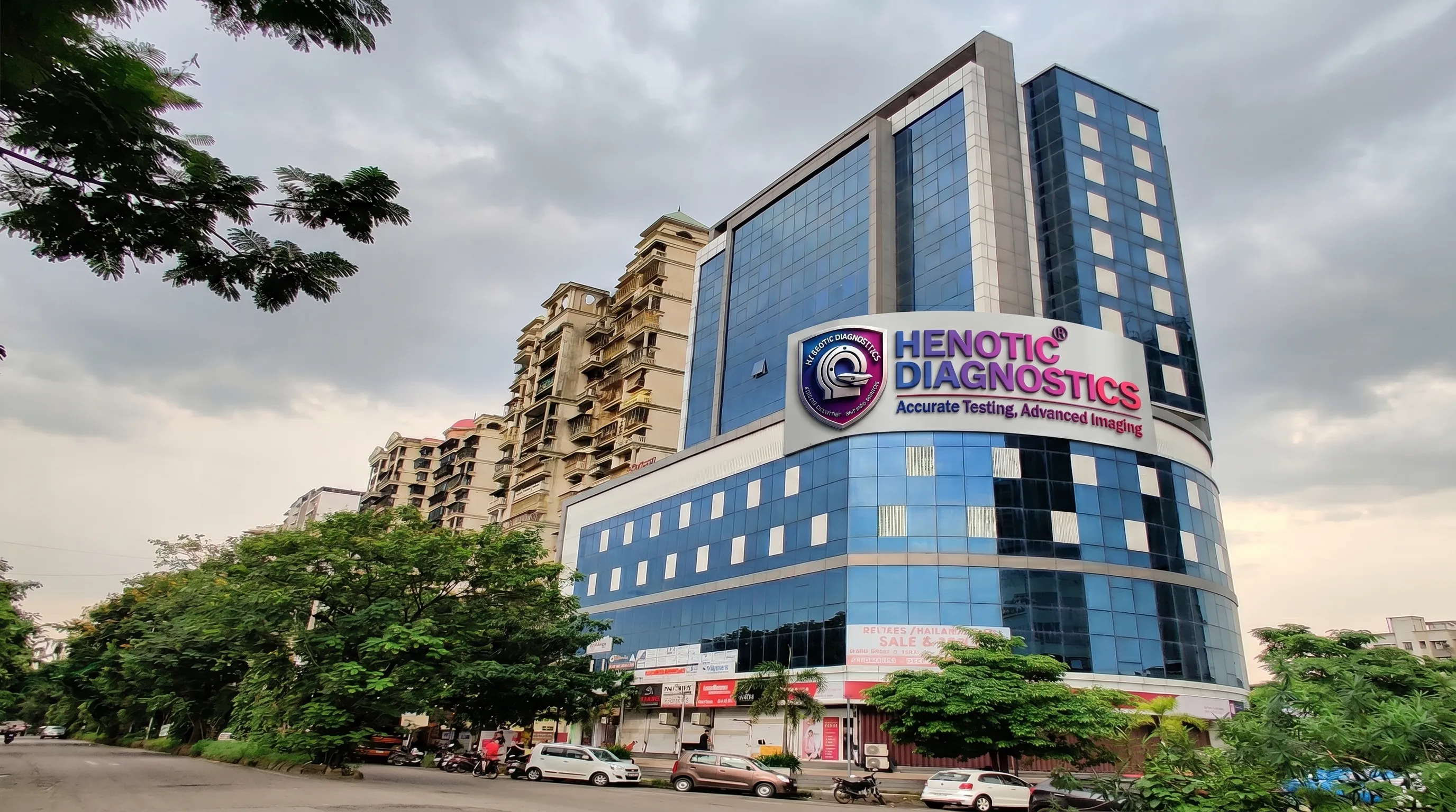
Henotic Diagnostics operates high-precision flagship and diagnostic network hubs near you, providing standardized clinical scans and pathology.
Main Flagship & Corporate Booking Hub
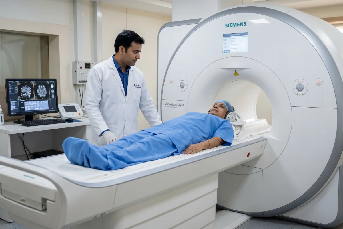
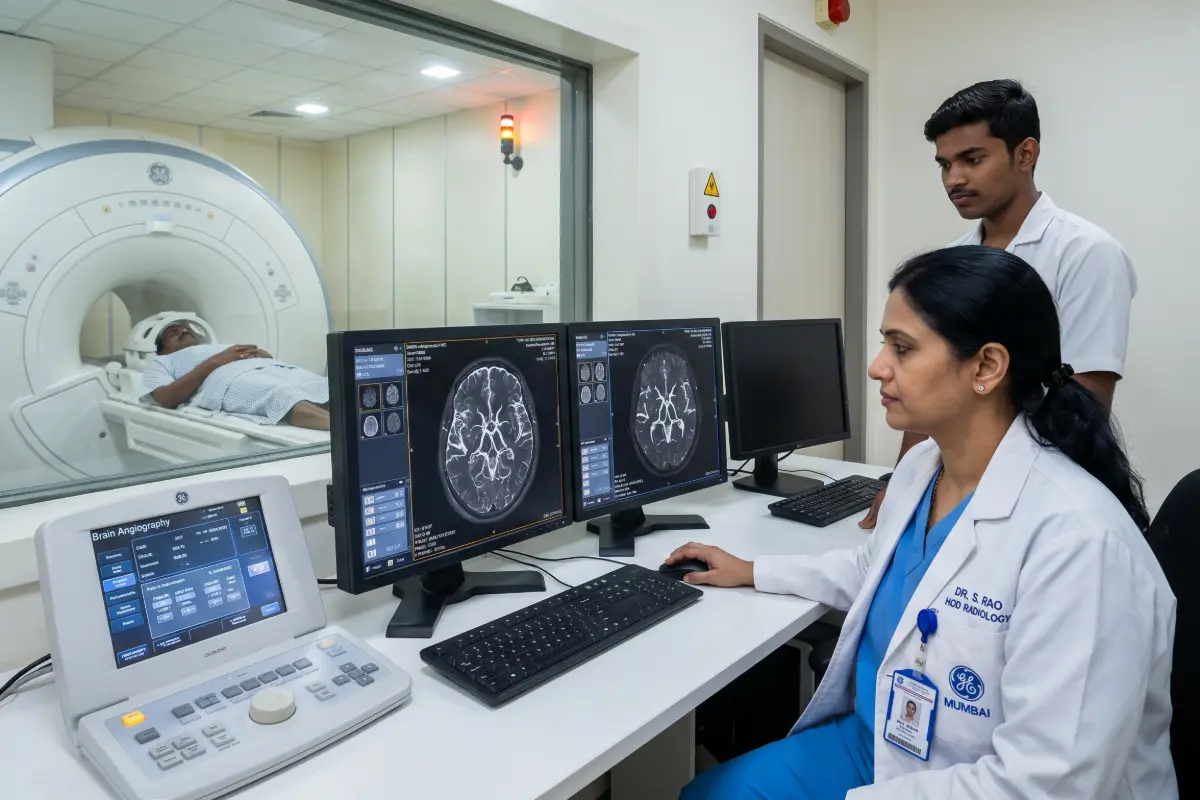
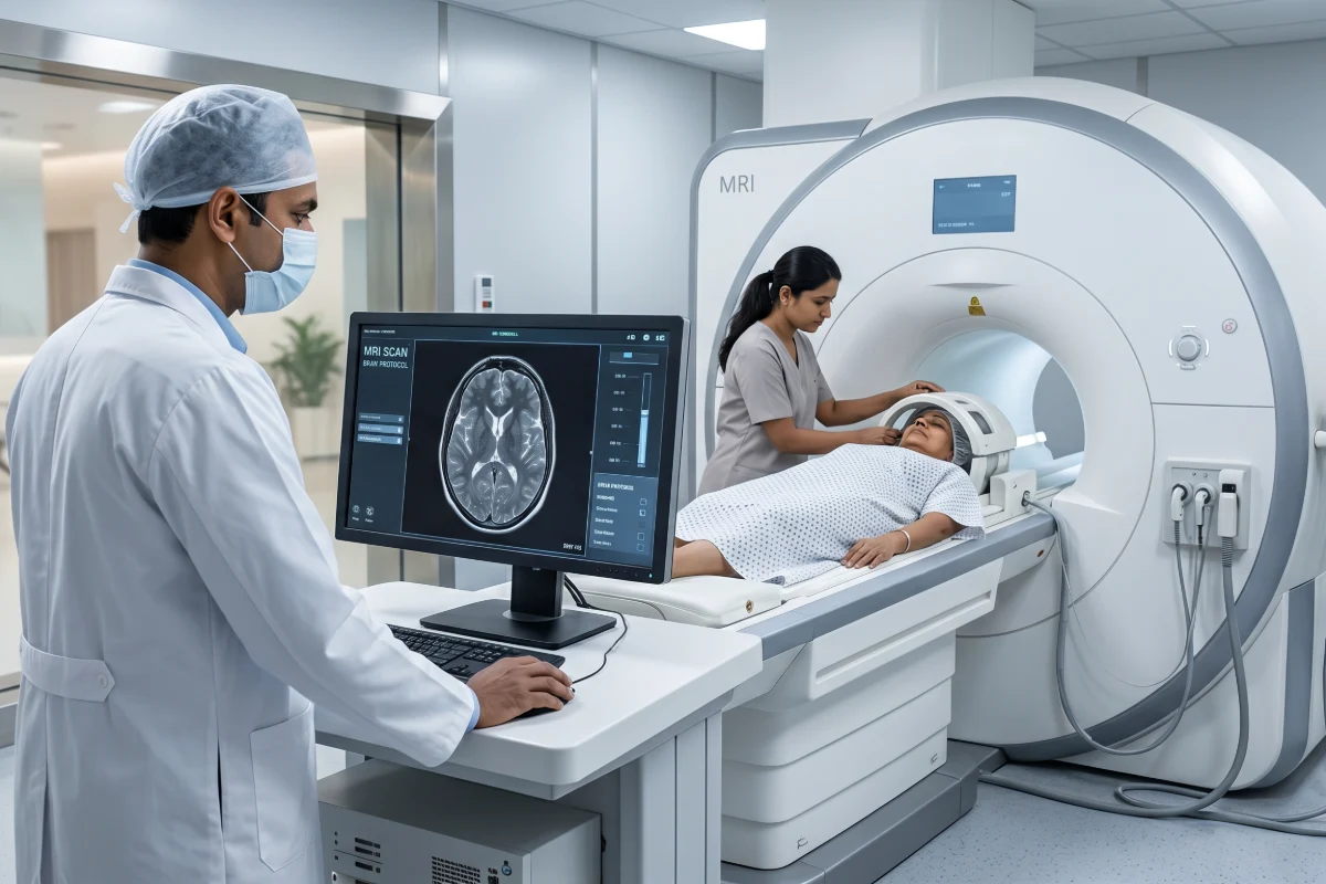
An MRI Pituitary Brain is a highly specialized magnetic resonance imaging examination designed to evaluate the pituitary gland, pituitary stalk, hypothalamus, optic chiasm, cavernous sinuses, and surrounding brain structures with exceptional anatomical detail. Specifically, by using advanced high-resolution MRI technology, this examination helps physicians diagnose pituitary tumors, endocrine disorders, hormonal abnormalities, congenital conditions, inflammatory diseases, vascular abnormalities, and postoperative changes without exposing patients to ionizing radiation.
Furthermore, because the pituitary gland controls numerous hormones responsible for growth, metabolism, reproduction, stress response, thyroid function, adrenal function, and water balance, accurate imaging plays a critical role in diagnosing disorders affecting the body’s endocrine system. Consequently, our dedicated protocols utilize thin imaging slices, specialized sequences, and dynamic contrast enhancement. As a result, radiologists easily identify even very small abnormalities that may not be visible on routine brain MRI examinations.
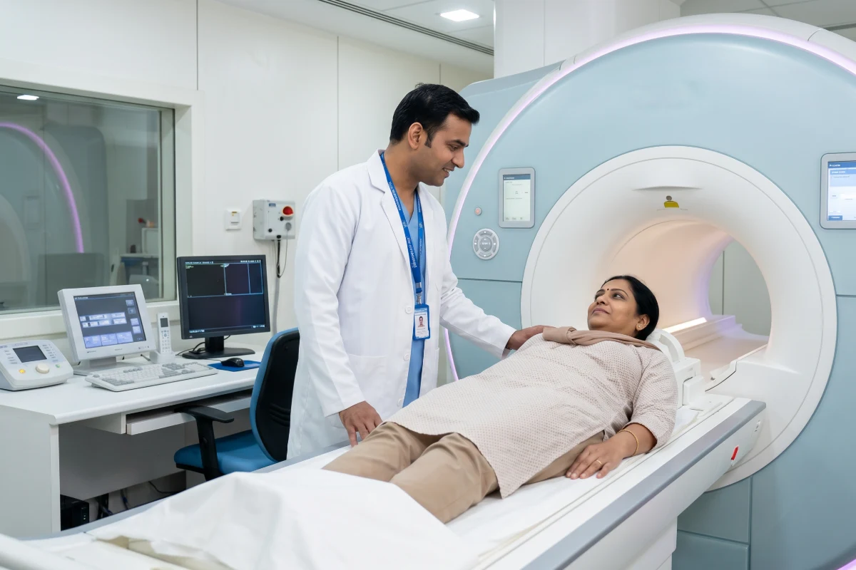
An MRI Pituitary Brain scan is a dedicated MRI examination that provides detailed multiplanar visualization of the pituitary gland and adjacent anatomical structures. Therefore, high-resolution imaging allows our radiologists to accurately evaluate gland size, symmetry, tissue characteristics, vascular relationships, enhancement patterns, and surrounding neurological structures for a comprehensive diagnostic assessment.
Physicians commonly recommend an MRI Pituitary Brain when they suspect disorders affecting hormone production, pituitary tumors, structural abnormalities, neurological symptoms, or visual disturbances related to pituitary enlargement. Moreover, the examination directly assists endocrinologists, neurologists, neurosurgeons, ophthalmologists, and oncologists in establishing accurate diagnoses and treatment strategies. Our experienced medical team ensures you receive precise results.
We evaluate gland size, morphology, enhancement characteristics, focal lesions, cystic abnormalities, and tissue integrity.
Our team assesses thickening, interruption, displacement, inflammatory disease, and congenital abnormalities.
The scan detects infiltrative disease, tumors, inflammatory disorders, developmental abnormalities, and structural changes.
We determine compression caused by pituitary macroadenomas or suprasellar masses affecting your vision.
The imaging evaluates tumor extension, vascular relationships, cranial nerve involvement, and invasion.
We assess the bony pituitary fossa for enlargement, remodeling, and structural abnormalities.
An MRI Pituitary Brain assists in diagnosing a wide range of endocrine, neurological, congenital, inflammatory, and neoplastic disorders affecting the pituitary gland and surrounding anatomical structures. You can learn more about related imaging standards from the RadiologyInfo guide on Head MRI.
If you experience subtle yet progressive signs, your doctor may suggest this scan. Similarly, if you require baseline laboratory testing for hormonal changes, you can review our blood test services.
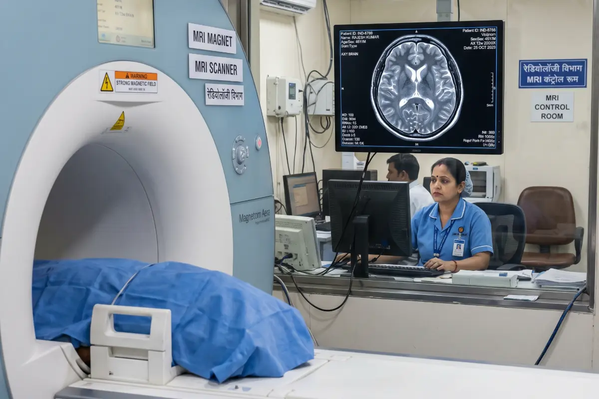
Additionally, a contrast-enhanced MRI Pituitary Brain significantly improves the visualization of pituitary lesions by highlighting differences in tissue vascularity and enhancement patterns. Dynamic contrast imaging proves particularly valuable for detecting pituitary microadenomas that may otherwise remain undetected.
The examination generally takes between 30 and 60 minutes depending on the imaging protocol and whether we perform contrast enhancement.
No. The examination is completely non-invasive and painless.
No. MRI uses powerful magnetic fields and radiofrequency waves instead of ionizing radiation.
Yes. Dedicated MRI Pituitary Brain protocols can identify lesions measuring only a few millimeters. You can read more regarding tumor detection from the Mayo Clinic.
Not always. However, contrast significantly improves detection of pituitary microadenomas and assessment of many pituitary disorders.
Yes. The scan clearly demonstrates the optic nerves, optic chiasm, and surrounding structures that may be affected by pituitary enlargement.
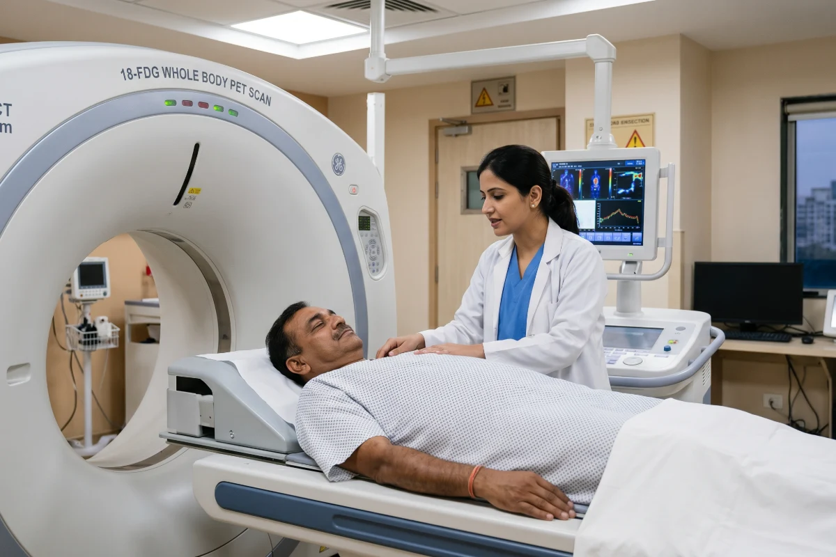
Ultimately, MRI offers superior soft tissue contrast, higher spatial resolution, multiplanar imaging capability, and improved detection of pituitary abnormalities compared with traditional CT imaging.
An MRI Pituitary Brain remains the imaging modality of choice for evaluating pituitary gland disorders because it provides exceptional visualization of endocrine anatomy. Furthermore, it accurately detects tumors of all sizes, assesses surrounding neurological structures, guides surgical planning, evaluates treatment response, and supports long-term patient care without exposing patients to ionizing radiation. Hence, its high diagnostic accuracy makes it indispensable in modern endocrine and neurological imaging.
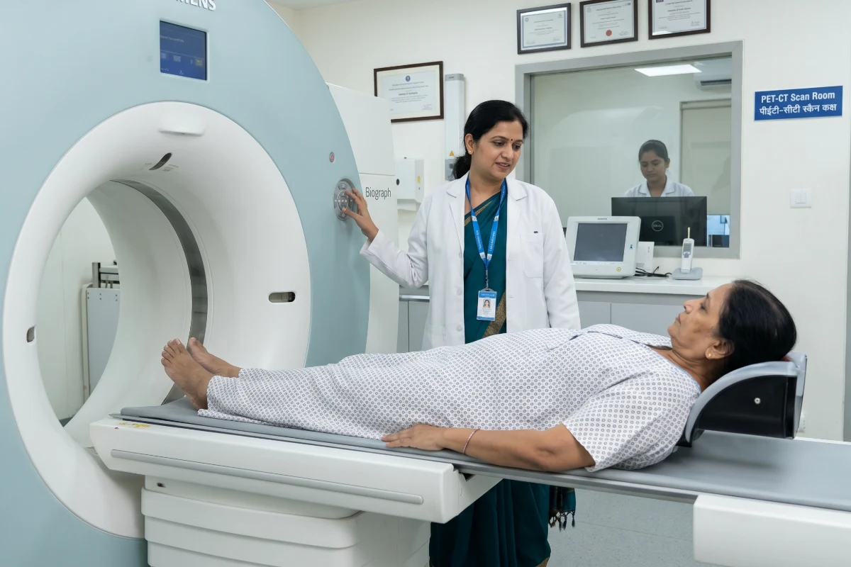
Starting @ High-resolution 3T MRI specifically configured with thin-slice imaging to map the tiny structures of the pituitary gland and hypothalamus. Starting @ Specialized dynamic contrast enhancement designed specifically to detect pituitary microadenomas that standard brain MRIs often miss. Starting @ Comprehensive evaluation of the sella turcica, optic chiasm, and cavernous sinuses to accurately diagnose hormonal imbalances. Starting @ Highly targeted imaging sequences focused entirely on the pituitary fossa to assess structural integrity, cysts, and tumors.

3T Pituitary MRI
₹8000 ₹1499
Salient Features
Dynamic Contrast MRI
₹9500 ₹1850
Salient Features
Premium Neuro-Endocrine
₹9000 ₹1600
Salient Features
Dedicated Protocol
₹8500 ₹1500
Salient Features
See what our patients have to say about their diagnostic experience with us.

"Excellent service! The staff is very cooperative and the facility is spotless. Got my MRI done smoothly."
"State-of-the-art equipment and highly professional doctors. Reports were delivered on time via WhatsApp. Highly recommended."
"Very convenient location in Kharghar. The home collection team was punctual and well-trained. Great experience overall."