Loading Diagnostics...
Connecting to our secure server
Connecting to our secure server
12 people booked MRI Scan
in Kharghar today • Verified

Access world-class diagnostic technology across our comprehensive network in Mumbai Suburban. Select your nearest neighborhood for priority booking.
Select your specific location to view tailored availability and book your slot.
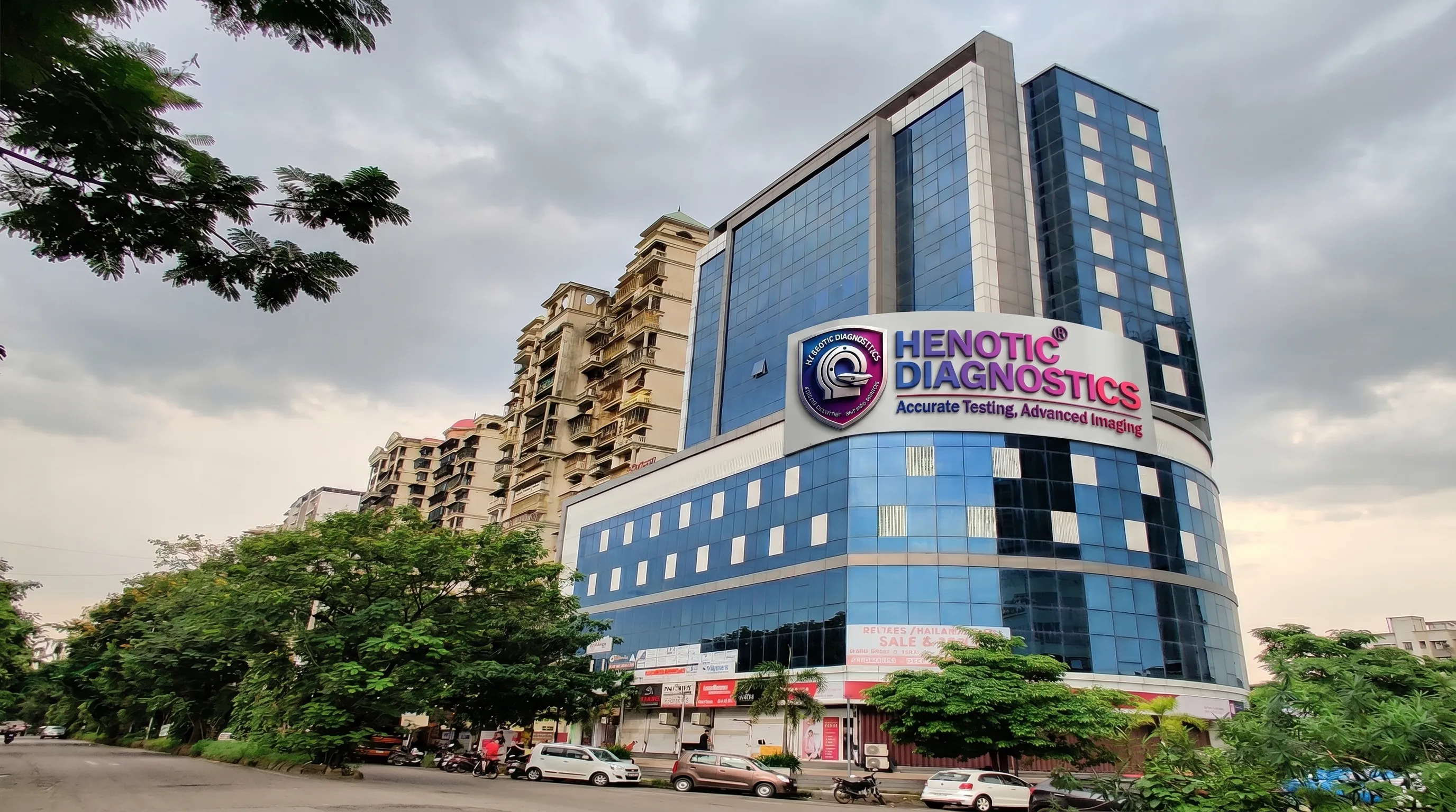
Henotic Diagnostics operates high-precision flagship and diagnostic network hubs near you, providing standardized clinical scans and pathology.
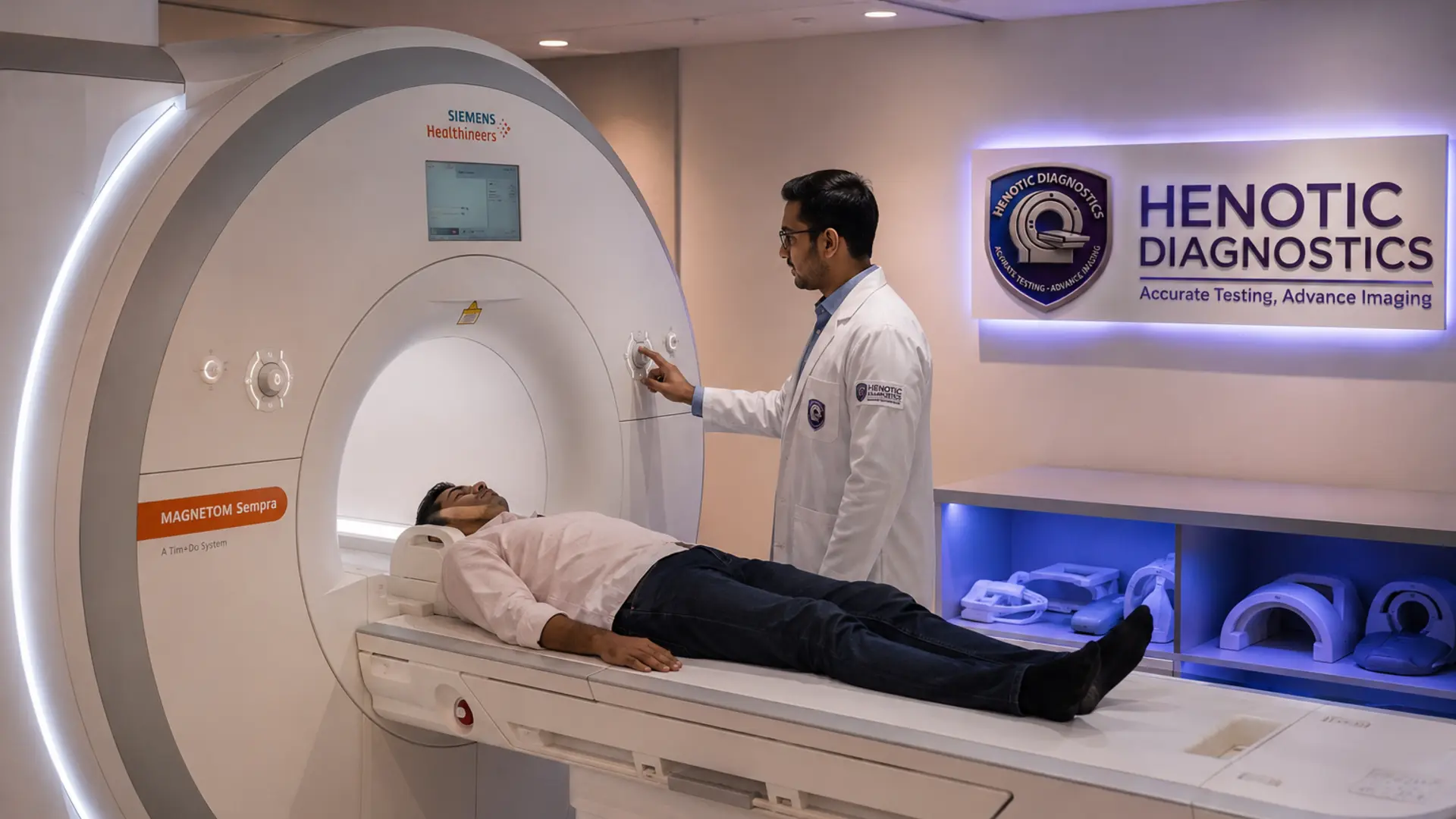
Main Flagship & Corporate Booking Hub
Experience the absolute highest standard of clinical clarity. Our cutting-edge, high-resolution joint neuroimaging technology ensures precise evaluation of chronic foot pain, complex ligament tears, Achilles tendon disorders, and hidden structural abnormalities. Consequently, you can confidently obtain rapid, reliable results to fast-track your recovery plan.
Trust Signals: NABL Quality Guidelines | Advanced 3.0T Technology | Expert Radiologists
WhatsApp Appointment | Call 088793 27184
An MRI Ankle scan is a non-invasive diagnostic technique that utilizes a remarkably powerful magnetic field and radiofrequency pulses. Consequently, this state-of-the-art system generates highly detailed multiplanar images across axial, coronal, and sagittal views without exposing you to harmful radiation.
Furthermore, physicians favor this imaging protocol whenever they suspect internal derangement. Unlike routine standard X-rays, magnetic resonance imaging provides exceptional soft tissue visualization. Meanwhile, it captures subtle variations within deep structures, which significantly enhances total diagnostic accuracy for medical specialists worldwide.
To understand why orthopedic surgeons prefer MRI, we must compare its capabilities against other standard imaging tests for lower limb soft tissues.
| Imaging Trait | MRI Scan | CT Scan | Ultrasound | Digital X-Ray |
|---|---|---|---|---|
| Soft Tissue Contrast | Excellent | Poor | Moderate | None |
| Bone Marrow Edema | Gold Standard | Invisible | Invisible | Invisible |
| Ionizing Radiation | None (Safe) | High Risk | None (Safe) | Low Risk |
| Deep Joint Cartilage | Highly Detailed | Limited Detail | Obstructed Views | Joint Space Only |
Our expert radiologists use this technology to map every intricate component of your lower joint.
The scan perfectly traces the cortical bone borders of the distal tibia and fibula. Additionally, it accurately screens the talus, calcaneus (heel bone), navicular, and adjacent cuboid components for micro-fractures.
It provides pristine images of the anterior talofibular ligament (ATFL), calcaneofibular ligament (CFL), and posterior talofibular ligament (PTFL). Similarly, it evaluates the deep medial deltoid ligament network.
The thick Achilles tendon complex remains highly visible throughout its insertion pathway. Moreover, it tracks the tibialis anterior, tibialis posterior, and localized peroneal tendons for structural alignment.
This comprehensive scan isolates exact structural failures. Therefore, it effectively identifies the following pathologies:
You should consult your orthopedic specialist and consider advanced imaging if you experience any of these persistent clinical manifestations:
Depending on your specific clinical history, your referring physician will request one of two primary approaches.
This approach represents the standard imaging sequence utilizing raw structural magnetic fields. Specifically, it accurately screens over 90% of routine orthopedic injuries, including sports sprains, tendon tears, and stress fractures.
Technologists inject an intravenous gadolinium element to heighten soft tissue visibility. Consequently, this hyper-vascular tracking is required to expose deep infectious borders (osteomyelitis), post-surgical scar tissue, and tumoral margins.
Following these simple steps ensures a safe, efficient, and comfortable imaging experience.
Once the scan completes, our consultant radiologist evaluates the sequences. Here is what common clinical terms in your report mean:
No. MRI utilizes powerful magnetic fields and radio waves, meaning it is 100% radiation-free and inherently safe for repeated diagnostic use.
A standard ankle scan requires approximately 20 to 35 minutes. However, if your doctor requests a contrast injection, the procedure may take up to 45 minutes.
Yes. High-resolution multiplanar imaging sequences clearly visualize nerve entrapment zones, deep neuromas, and conditions like tarsal tunnel syndrome.
While MRI involves no radiation, standard medical protocol advises avoiding non-emergency scans during the first trimester. Always consult your obstetrician prior to booking.
Our expert radiologists process and deliver comprehensive, cross-verified structural digital reports within 24 hours of scan completion.
According to recent clinical evaluations published by organizations such as the National Institutes of Health (NIH), magnetic resonance profiling exhibits a diagnostic sensitivity above 95% for acute ligamentous trauma. Consequently, it remains the diagnostic gold standard for evaluating complex sports injuries and joint pathologies globally.
“Early MRI diagnosis of ankle trauma significantly reduces the risk of chronic joint instability by guiding precise, conservative, or surgical treatment plans.”
See what our patients have to say about their diagnostic experience with us.

"Excellent service! The staff is very cooperative and the facility is spotless. Got my MRI done smoothly."
"State-of-the-art equipment and highly professional doctors. Reports were delivered on time via WhatsApp. Highly recommended."
"Very convenient location in Kharghar. The home collection team was punctual and well-trained. Great experience overall."