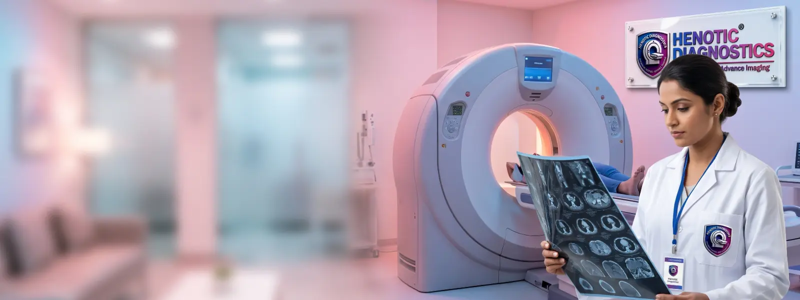
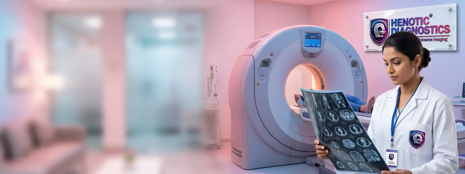
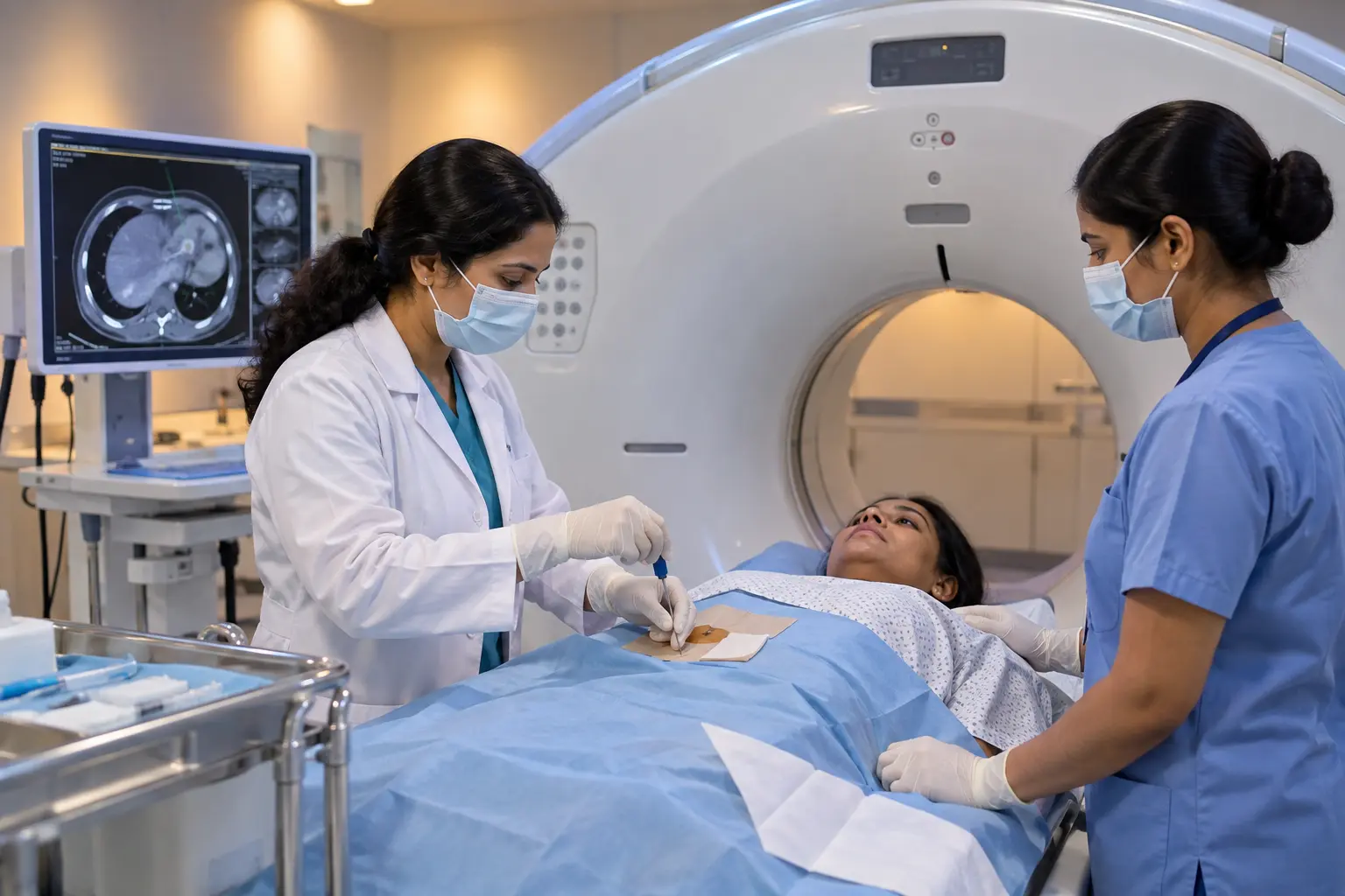
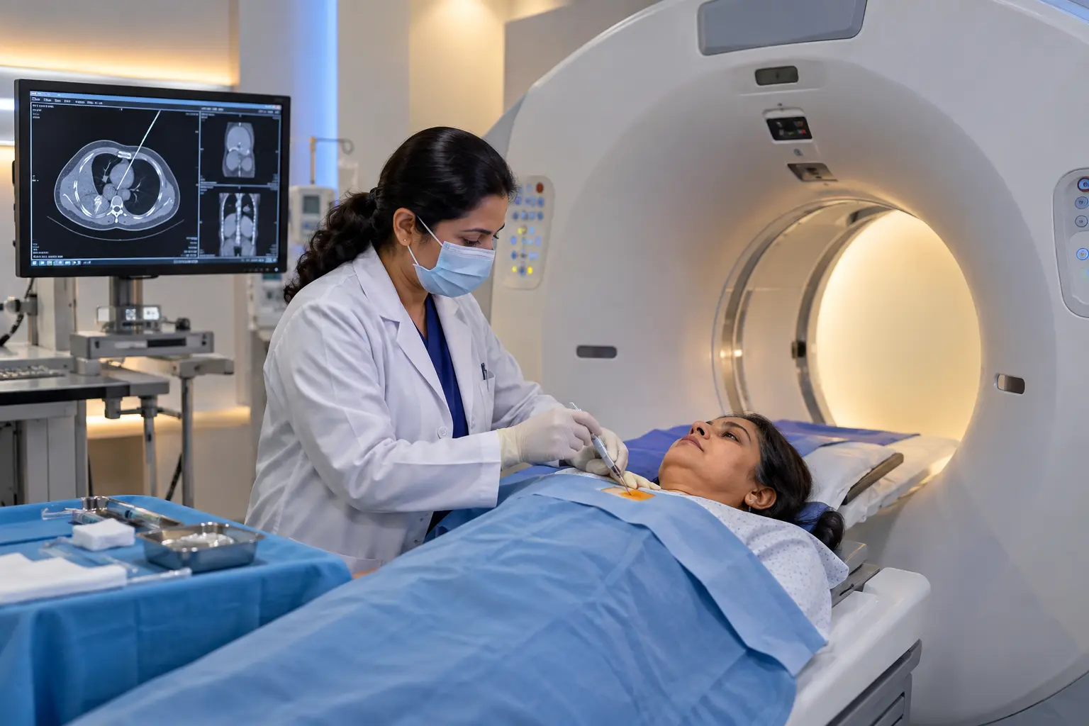
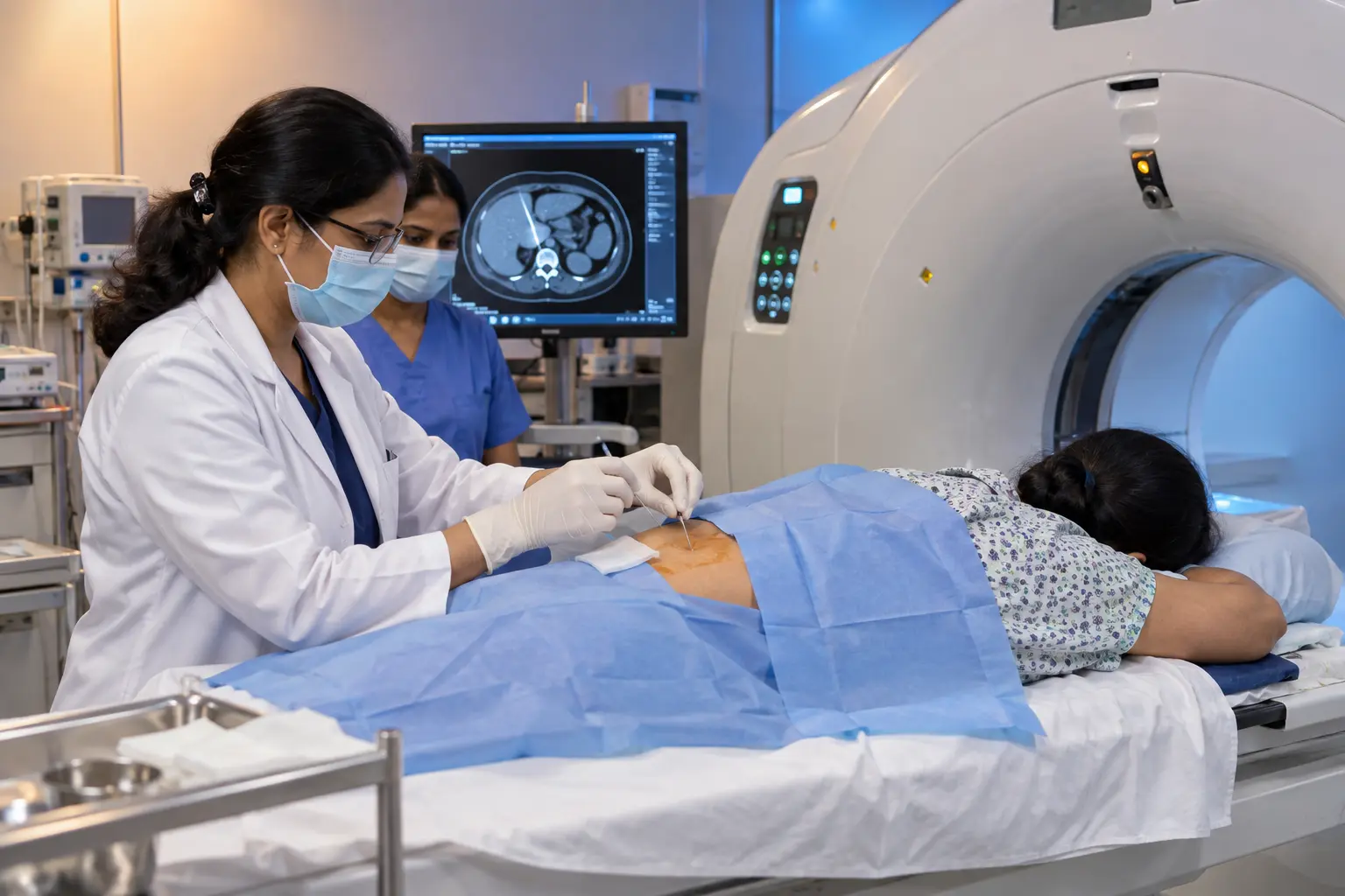
CT Guided FNAC diagnostic procedure suite at Henotic Diagnostics
What Is CT Guided FNAC?
Understanding Percutaneous Needle Aspiration
CT Guided FNAC (Computed Tomography Guided Fine Needle Aspiration Cytology) is an image-guided diagnostic procedure. Specifically, this procedure uses cross-sectional CT scanning to direct a fine needle into a targeted mass, nodule, or selected lesion. Therefore, the primary goal of CT guided fine needle aspiration is to collect cellular material or fluid aspirate for cytological evaluation in a pathology laboratory. At Henotic Diagnostics Kharghar, our radiologists choose CT guidance when target lesions lie deep inside the body. Furthermore, radiologists utilize CT scans whenever thick anatomical structures obscure lesions or whenever ultrasound cannot visualize the tissue clearly. Consequently, CT imaging provides precise cross-sectional localization to assist clinical diagnostic pathways. For further details on percutaneous diagnostic procedures, patients can consult the RadiologyInfo Percutaneous Biopsy Guide.
Why Is CT Guidance Used for Fine Needle Aspiration?
The choice of imaging guidance depends on the lesion location, visibility, accessibility, surrounding anatomy, clinical requirements, and procedural safety. While doctors frequently choose ultrasound for superficial structures like thyroid nodules, CT Guided FNAC provides distinct anatomical advantages for deep body regions. In addition, radiologists rely on CT guidance for complex anatomical structures because cross-sectional scans deliver precise clarity.
Key Anatomical Advantages of CT Guidance
Furthermore, CT imaging offers several key benefits during needle aspiration:
Cross-Sectional Anatomical Detail: First, CT scans clearly delineate lesions situated deep within the chest, abdomen, pelvis, or retroperitoneum.
Visualization Unhindered by Gas or Bone: Second, CT imaging provides clear cross-sectional visualization in regions where depth, bone structure, or intestinal gas limits ultrasound visibility.
Needle Trajectory Planning: Third, serial CT planning scans confirm exact needle placement in three dimensions before the radiologist aspirates cellular material.
Vascular and Visceral Path Mapping: Finally, cross-sectional views allow the operating radiologist to map an optimal access path around major blood vessels and organs.
When Is CT Guided FNAC Recommended?
Physicians usually recommend CT Guided FNAC when prior diagnostic imaging identifies an indeterminate mass or suspicious nodule. For instance, an abdominal CT, chest CT, or PET-CT scan may reveal deep lesions requiring cellular evaluation. Consequently, doctors request image-guided fine needle aspiration when a lesion is non-palpable or when sonographic visualization encounters physical obstacles. Moreover, practice parameters from the American College of Radiology (ACR) emphasize selecting image guidance based on spatial accessibility and patient safety.
What Lesions Can Be Sampled?
Depending on clinical indication, lesion accessibility, and patient anatomy, radiologists utilize computed tomography guidance to evaluate target lesions across various anatomical regions. In addition, treating physicians rely on this technique to gather cellular diagnostic data safely.
Anatomical Regions Suitable for Diagnostic Sampling
Specifically, radiologists frequently evaluate the following targeted areas:
Selected Thoracic & Mediastinal Lesions: First, CT scans help sample deep mediastinal masses or pulmonary nodules located near chest wall structures.
Retroperitoneal Masses & Lymph Nodes: Second, CT guidance enables access to deep retroperitoneal lymphadenopathy, para-aortic masses, or adrenal lesions.
Abdominal Visceral Lesions: Third, radiologists target deep liver, pancreatic, or renal lesions previously identified on cross-sectional imaging.
Deep Pelvic & Soft Tissue Masses: Finally, CT guidance assists in sampling presacral, pelvic sidewall, or deep soft-tissue masses.
Note: Therefore, the treating clinical team determines suitability for CT guided fine needle aspiration on a case-by-case basis after reviewing prior diagnostic scans.
How Is CT Guided FNAC Performed?
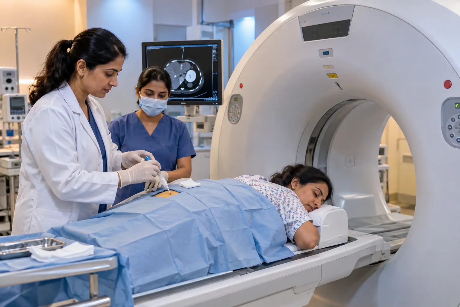
CT guided fine needle aspiration workflow and scanner setup
Radiologists conduct CT Guided FNAC following a structured diagnostic workflow designed for patient comfort and precision. In addition, our team adheres to strict clinical safety protocols throughout the procedure.
Phase 1: Localization & Preparation
Step 1 — Review of Prior Imaging: First, the radiologist reviews previous CT, MRI, or PET-CT scans to verify lesion location and clinical details.
Step 2 — Patient Positioning: Next, the clinical team positions the patient comfortably on the CT scanner table in a supine, prone, or lateral position.
Step 3 — CT Localization Scan: Then, the radiologist acquires initial CT localization images to map the exact entry trajectory and measure lesion depth.
Step 4 — Skin Preparation & Local Anesthesia: Subsequently, the staff cleans the skin with antiseptic solution, and the radiologist administers local anesthesia to numb superficial tissues.
Phase 2: Sampling & Analysis
Step 5 — Guided Needle Advancement: After numbing the area, the radiologist advances a fine needle toward the target. Meanwhile, intermittent CT scans verify the exact needle tip position.
Step 6 — Aspiration: Once in position, the radiologist applies gentle suction to collect cellular material or fluid aspirate from the lesion.
Step 7 — Specimen Preparation: Immediately afterward, the technician prepares the aspirated cells on microscope slides or places them in fixative liquid media for pathological analysis.
Step 8 — Post-Procedure Observation: Finally, our medical staff monitors the patient during a short post-procedure observation period to ensure safety before discharge.
What Preparation Is Required for CT Guided FNAC?
Preparation guidelines for CT guided fine needle aspiration vary depending on the target body region and clinical requirements. However, following standard instructions ensures a smooth and safe diagnostic experience.
Key Preparation Steps Before Your Test
Medical History & Medications: First, inform your doctor about all current medications you take. Specifically, disclose blood thinners or anticoagulants, because your clinical team may adjust dosage schedules before the procedure.
Previous Imaging Reports: Second, bring all recent CT, MRI, USG, and laboratory reports to your appointment. Consequently, our radiologist can plan your procedure with complete clinical context.
Fasting Instructions: Third, follow any specific fasting instructions provided by your clinical team. Fasting requirements vary depending on contrast media usage or sedation protocols.
Allergies & Medical Conditions: Finally, notify our staff if you have allergies to local anesthetics, iodine contrast media, or latex. As a result, we can take proper precautionary measures.
Is CT Guided FNAC Painful?
To keep patients comfortable, the radiologist administers local anesthesia to numb the skin and tissue along the needle path. Consequently, most patients feel minimal pain during CT Guided FNAC. However, patients may experience brief pressure or mild soreness depending on the lesion site. Overall, individual experiences vary based on procedural complexity, but local numbing effectively minimizes discomfort.
Does CT Guided FNAC Use Radiation?
Yes, CT Guided FNAC utilizes low-dose ionizing radiation scans. Specifically, the radiologist performs brief CT acquisitions to map lesion positioning and confirm needle placement. Furthermore, our clinical team optimizes every scan protocol in accordance with ALARA (As Low As Reasonably Achievable) radiation safety guidelines. Therefore, we ensure maximum diagnostic accuracy while keeping radiation exposure as low as possible.
What Happens to the Diagnostic Sample?
The material collected during fine needle aspiration consists of loose cellular clusters, individual cells, or fluid aspirate. Immediately following aspiration, our laboratory team initiates detailed cytopathological processing.
Laboratory Cytology Processing Workflow
In addition, pathologists evaluate the specimen using specialized laboratory steps:
Slide Preparation: First, technicians prepare cellular smears on microscope slides and preserve them using air-drying or alcohol fixation techniques.
Liquid-Based Cytology: Second, the technician places needle rinses into specialized fixative solutions for automated cell processing.
Microscopic Examination & Diagnostic Interpretation: Finally, a cytopathologist examines the stained cells under high magnification to analyze cellular features. Because fine needle aspiration evaluates individual cells rather than intact tissue architecture, your doctor may order a core biopsy if detailed structural evaluation is necessary.
Both CT Guided FNAC and CT guided core biopsy serve as valuable diagnostic sampling techniques. However, they collect different specimen types for distinct laboratory evaluations. Therefore, understanding their differences helps clarify why a physician chooses one method over another.
Comparative Analysis of Diagnostic Sampling Procedures
Comparison: CT Guided FNAC vs Core Biopsy
Feature
CT Guided FNAC
CT Guided Core Biopsy
Sampling Method
Fine needle aspiration
Core needle biopsy
Specimen Collected
Cells, cellular material, or fluid aspirate
Intact tissue core specimen
Main Laboratory Evaluation
Cytology / Cytopathology
Histopathology
Tissue Architecture
Limited structural architecture
Preserved intact tissue architecture
Selection
Depends on clinical question
Depends on clinical question
Consequently, the treating clinical team selects the appropriate sampling procedure based on lesion location, suspected pathology, and necessary diagnostic details.
CT Guided FNAC vs Ultrasound-Guided FNAC
The choice of imaging guidance depends on lesion visibility, depth, accessibility, and surrounding anatomical structures. While neither modality is universally superior, doctors select each modality based on specific clinical requirements. For instance, CT Guided FNAC excels in deep or complex regions where ultrasound encounters physical limitations.
Comparing Guidance Modalities
Comparison: CT Guidance vs Ultrasound Guidance
Parameter
CT Guided FNAC
Ultrasound-Guided FNAC
Imaging Method
Cross-sectional Computed Tomography scans
Real-time sonographic acoustic waves
Typical Usefulness
Selected deep, retroperitoneal, thoracic, pelvic, or bone/gas-obscured lesions
In addition, radiologists frequently prefer CT guidance when intestinal gas or bony structures obscure ultrasound beams.
Possible Risks and Limitations of CT Guided FNAC
Procedural risks vary according to target lesion location, access path, and individual patient health. However, CT Guided FNAC remains a safe and well-tolerated procedure when performed by experienced radiologists.
Understanding Potential Procedural Risks
Furthermore, patients should understand the following potential risks and limitations:
Local Discomfort & Soreness: First, patients may experience mild tenderness or soreness near the needle entry site.
Minor Bleeding or Bruising: Second, minor localized bleeding or small hematomas can develop along the needle path.
Infection: Third, a minimal risk of localized skin or tissue infection exists, though sterile techniques mitigate this risk.
Non-Diagnostic Sample: Fourth, an aspirate may contain sparse cells, fluid only, or necrotic material. Consequently, a non-diagnostic sample does NOT mean the lesion is benign; rather, it indicates that the sample lacks sufficient cellular details for interpretation.
Procedure-Specific Complications: In addition, thoracic procedures carry a minor risk of pneumothorax or mild hemoptysis depending on lesion location.
Diagnostic Limitations: Finally, if cytology results remain inconclusive, your doctor may recommend repeat sampling, core biopsy, or follow-up imaging.
How Are CT Guided FNAC Results Reported?
Immediately following specimen collection, our courier delivers the slides and cell suspensions directly to the pathology laboratory. Afterwards, a cytopathologist evaluates the cellular features to distinguish between benign, suspicious, and malignant conditions.
Diagnostic Report Categories
Generally, pathologists classify diagnostic findings into three main categories:
Diagnostic: First, a diagnostic result means the specimen contains sufficient cellular material for a definitive cytological interpretation.
Indeterminate / Inconclusive: Second, an indeterminate result indicates atypical cellular features that require additional imaging or tissue sampling.
Non-Diagnostic / Inadequate: Third, a non-diagnostic result means the aspirate lacks adequate cellular material for evaluation.
Consequently, if a sample proves non-diagnostic, your treating physician may request clinical correlation, repeat sampling, core biopsy, or follow-up imaging. Therefore, your doctor will interpret these laboratory findings alongside your complete diagnostic context.
Frequently Asked Questions About CT Guided FNAC
General Procedure FAQs
What is CT Guided FNAC?
CT Guided FNAC (Fine Needle Aspiration Cytology) is an image-guided diagnostic procedure. Specifically, radiologists use cross-sectional computed tomography imaging to visualize a target lesion and guide a fine needle to collect cellular material or fluid for microscopic examination.
Why is CT used for FNAC?
Radiologists choose CT guidance when a lesion lies deep within the body, behind bone structures, or inside gas-filled tissue. Consequently, CT imaging provides clear visualization where sonography encounters physical limitations.
How is CT Guided FNAC performed?
First, the clinical team positions the patient on the CT table and acquires localization scans. Next, the radiologist cleans the skin and applies local anesthesia. Then, under CT guidance, the radiologist advances a fine needle into the target area and aspirates cellular material for laboratory analysis.
Is CT Guided FNAC painful?
To ensure patient comfort, the radiologist applies local anesthesia to numb the skin and superficial tissue. Therefore, patients usually feel only brief pressure or mild soreness during the procedure.
Does CT Guided FNAC use radiation?
Yes, CT Guided FNAC uses ionizing radiation scans. However, our team optimizes scan acquisitions according to radiation safety principles such as ALARA to minimize exposure.
Preparation and Diagnostic Results FAQs
Does CT Guided FNAC require fasting?
Fasting requirements depend on the target lesion location, intravenous contrast requirements, and sedation plans. Therefore, patients should follow the specific preparation guidelines provided by their clinical team.
What does FNAC collect?
Fine needle aspiration collects loose cellular material, small cell clusters, or fluid aspirate from a target lesion for detailed cytopathological evaluation.
What happens if FNAC is non-diagnostic?
A non-diagnostic sample indicates that the aspirate lacks sufficient cellular material for a definitive answer. Consequently, your physician may recommend repeat sampling, a core biopsy, or follow-up imaging.
What is the difference between FNAC and core biopsy?
Fine needle aspiration cytology uses a thin needle to collect loose cellular material for cytology. In contrast, a core needle biopsy uses a larger needle to extract an intact tissue core for histopathology.
What is the difference between CT Guided FNAC and ultrasound-guided FNAC?
Ultrasound-guided FNAC uses real-time sonography and works best for superficial lesions like thyroid nodules. In contrast, computed tomography guidance is selected when depth, bone, or intestinal air limits ultrasound visibility.
At Henotic Diagnostics Kharghar, experienced radiologists supervise all diagnostic cross-sectional imaging procedures. Specifically, Dr. Pratibha Patil brings over 16 years of clinical radiology experience in diagnostic scanning and interventional guidance. Consequently, she works closely with referring physicians to deliver accurate cellular diagnostic reports.
Quality Standards & Diagnostic Accreditations
PCPNDT Certified
NABL Accredited
ISUOG Member
Visit Henotic Diagnostics
Henotic Diagnostics in Kharghar provides advanced cross-sectional imaging and diagnostic evaluation services. Furthermore, our clinical team prioritizes patient comfort, strict safety protocols, transparent counseling, and prompt reporting.
Address: Second floor, Millennium Empire, Business Park, Plot No 47, D Mart Rd, Sector 15, Kharghar, Navi Mumbai, Panvel, Maharashtra 410210.
If your doctor has recommended CT Guided FNAC or diagnostic cross-sectional imaging, please contact our scheduling desk today. Furthermore, our dedicated patient care team is ready to assist you with appointment bookings and pre-procedure guidance.
You can book your CT Guided FNAC appointment at our Kalamboli center through multiple convenient channels: call us directly at 088793 27184, send a WhatsApp message for instant booking, or use the online booking form on our website. Walk-in appointments are also welcome based on slot availability. We recommend advance booking to ensure zero wait times.
Yes, Henotic Diagnostics Kalamboli is empanelled with all major insurance companies and TPAs (Third Party Administrators) including Star Health, ICICI Lombard, New India Assurance, HDFC Ergo, Max Bupa, Bajaj Allianz, and many more. We offer cashless processing for CT Guided FNAC where applicable. Please carry your insurance card and a valid ID for seamless processing.
At Henotic Diagnostics Kalamboli, most CT Guided FNAC reports are delivered within 2-6 hours for routine tests. Critical and urgent reports are expedited on a priority basis. All reports are delivered digitally via WhatsApp, email, and our secure online patient portal. Hard copies are also available at the center upon request. You will receive an SMS/WhatsApp notification the moment your report is ready.
Henotic Diagnostics Kalamboli operates 24 hours a day, 7 days a week, including Sundays and public holidays. This means you can schedule your CT Guided FNAC at a time that is most convenient for you — early morning, late evening, or even overnight for emergency requirements. Our radiology and pathology departments maintain round-the-clock availability.
Yes, Henotic Diagnostics Kalamboli is fully accredited by NABL (National Accreditation Board for Testing and Calibration Laboratories) and ISO 9001:2015 certified. Our radiology equipment also complies with AERB (Atomic Energy Regulatory Board) safety standards. These accreditations ensure that every CT Guided FNAC performed at our facility meets the highest national and international quality benchmarks for accuracy, safety, and reliability.
The cost of CT Guided FNAC at Henotic Diagnostics Kalamboli starts from ₹2,000-2,500 for plain scans, which is 40-50% lower than market rates of ₹4,000-6,000. Contrast-enhanced CT scans and specialized studies like CT angiography may have different pricing. We offer transparent, upfront pricing with no hidden fees. Call us or WhatsApp for the exact cost based on your doctor's prescription.
Preparation for CT Guided FNAC depends on the type of scan: (1) Plain CT Scan — generally no preparation needed. (2) Contrast CT — fasting for 4-6 hours is required; a kidney function test (serum creatinine) within the last 3 months is mandatory. (3) CT Abdomen — you may be asked to drink oral contrast 1-2 hours before the scan. (4) Inform your doctor about any allergies, especially to iodine-based dyes. (5) Bring your prescription, previous reports, and insurance card.
A CT Guided FNAC at Henotic Diagnostics Kalamboli is extremely quick — the actual scan takes only 5-15 minutes depending on the body region. Our advanced 128-slice CT scanner captures high-resolution images in seconds. Including preparation time and contrast injection (if applicable), the entire process typically takes 20-30 minutes. Reports are usually available within 2-4 hours.
Modern CT scanners at Henotic Diagnostics use advanced dose-reduction technology (ALARA principle) to minimize radiation exposure while maintaining image quality. A single CT scan uses a very small amount of radiation that is generally considered safe for diagnostic purposes. However, CT scans are not routinely recommended for pregnant women. Our AERB-certified equipment and protocols ensure that radiation doses are kept as low as reasonably achievable.
CT scans use X-rays and are excellent for imaging bones, lungs, and acute emergencies (like bleeding or fractures). They are very fast (seconds to minutes). MRI uses magnetic fields and radio waves (no radiation) and provides superior soft tissue detail — ideal for brain, spine, joints, and ligaments. Your doctor will recommend the appropriate modality based on your clinical condition. At Henotic Diagnostics Kalamboli, we offer both with expert radiologist interpretation.
Yes, a valid doctor's prescription or referral is required for a CT Guided FNAC at Henotic Diagnostics Kalamboli. This ensures that the correct scan protocol is used for your clinical condition, the appropriate body part is scanned, and the radiologist can provide a meaningful clinical correlation in the report. If you need a consultation, we can connect you with our in-house physicians.
Local FAQs
CT Guided FNAC in Kalamboli
Answers to the most common questions about CT Guided FNAC services near Kalamboli.
Yes, Henotic Diagnostics offers CT Guided FNAC services near Kalamboli, Navi Mumbai. Our NABL-accredited centre provides accurate diagnostics with state-of-the-art equipment and experienced radiologists. Book your appointment today for priority service.
The cost of CT Guided FNAC at Henotic Diagnostics starts from competitive rates. We offer up to 50% savings compared to market rates in Kalamboli, with no compromise on quality. Contact us at +91 8879327184 for the latest pricing.
You can book CT Guided FNAC near Kalamboli by calling +91 8879327184 or booking online through our website. We offer instant confirmation, zero waiting time with prior booking, and WhatsApp report delivery for your convenience.
Yes, most CT Guided FNAC reports at Henotic Diagnostics are available within 24 hours, with many delivered same day. Reports are shared digitally via WhatsApp and email, so patients in Kalamboli can access results quickly without revisiting the centre.
Also Available In Navi Mumbai
Get Ct Guided Fnac at other Henotic Diagnostics centers near you
Henotic Diagnostics offers affordable Ct Guided Fnac services across Navi Mumbai — including Vashi, Sanpada, Juinagar, Nerul, Seawoods, and 10 more locations. All centers feature NABL-accredited labs, AERB-certified equipment, and same-day digital report delivery.
If you are looking for a highly accurate Ct Guided Fnac near Kalamboli, Henotic Diagnostics is your trusted partner. Easily accessible from all major points in Navi Mumbai, our world-class facility combines highly experienced radiologists with state-of-the-art medical technology. Patients from Kalamboli consistently rate us 4.7/5 Stars (based on 290 reviews) for our compassionate care, zero wait times, and instant report delivery.
Serving Micro-neighborhoods & Sectors:
Sector 1E KalamboliKalamboli CircleSteel Market RoadRoadpali
Frequently Asked Questions
How much does a Ct Guided Fnac cost in Kalamboli?+
The cost of a Ct Guided Fnac at our Kalamboli diagnostic center is highly competitive. We offer transparent pricing with zero hidden fees. Contact us directly for today's discounted rate.
How quickly can I get my Ct Guided Fnac reports in Navi Mumbai?+
For patients visiting from Kalamboli and across Navi Mumbai, Henotic Diagnostics guarantees fast turnaround times. Most Ct Guided Fnac reports are available the same day via secure WhatsApp delivery.
Is the Kalamboli center NABL & AERB accredited?+
Yes! Our entire facility serving Kalamboli maintains strict NABL, ISO, and AERB accreditations to guarantee 100% precision for your Ct Guided Fnac.