Loading Diagnostics...
Connecting to our secure server
Connecting to our secure server


Browse our comprehensive library of diagnostic capabilities. We provide over 200 NABL accredited radiology scans and automated clinical pathology investigations.
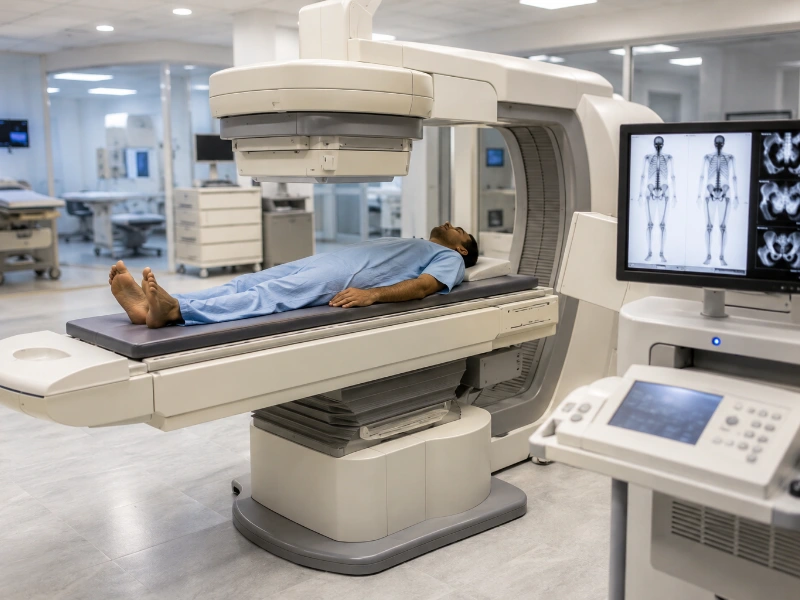
A Bone Scintigraphy Scan, commonly known as a nuclear bone scan, is a highly specialized diagnostic imaging procedure designed to monitor cellular metabolism and bone turnover across your entire skeletal network. Unlike traditional structural imaging methods like X-rays that only capture structural changes after significant mineral loss has occurred, bone scintigraphy measures the live biological activity of bone tissue. Specifically, by utilizing advanced dual-head gamma cameras and safe, bone-seeking radiotracers, our nuclear medicine department identifies micro-structural variations, stress fractures, infections, and oncological cell drops with exceptional diagnostic precision.
Furthermore, because your bone cells naturally increase their metabolic activity to repair hidden areas of stress, irritation, or disease, tracking these cellular changes offers an invaluable early window into your skeletal health. A dynamic bone issue can build silently long before a standard scan reveals a clear anatomical change, which makes functional tracking essential. Consequently, our dedicated diagnostic protocols employ ultra-sensitive gamma tracking crystals and standardized regional imaging to evaluate skeletal changes early. As a result, orthopedicians, oncologists, and rheumatologists can easily spot early stress fields, confirm underlying tissue joint disease, and guide highly effective care plans.
Bone scintigraphy is a functional molecular imaging profile that relies on the natural absorption properties of technetium-labeled diphosphonate compounds. Therefore, precise multi-tiered laboratory calibration allows our nuclear medicine physicians to accurately trace systemic cellular updates, locate high-turnover hot spots, and distinguish structural cell issues for a complete health assessment.
To ensure complete financial transparency for our patients, we provide clear, upfront cost structures across our advanced nuclear medicine imaging services. Below is the current price breakdown for bone imaging variations and associated biochemical panel matchings at our diagnostic hubs:
| Diagnostic Scan / Panel Variation | Estimated Cost (INR) |
|---|---|
| Standard Whole-Body Bone Scan | ₹5,000 – ₹6,500 |
| Three-Phase Bone Scintigraphy Scan | ₹6,500 – ₹8,000 |
| Advanced SPECT-CT Bone Scan | ₹8,500 – ₹11,000 |
Note: Final pricing variations can occur based on specialized pharmaceutical requirements or specific clinical instructions provided by your referring consultant.
Physicians commonly recommend a whole-body bone scan when investigating unexplained, continuous skeletal pain that standard structural checks cannot clear up, or when monitoring high-risk tissue recovery. Moreover, the analytical findings directly assist orthopedicians, oncologists, rheumatologists, and sports medicine specialists in choosing the safest treatment paths. Our experienced clinical team ensures you receive highly accurate imaging and reports.
Immediately following the tracer injection, our advanced gamma camera captures fast, sequential images of the target area. This dynamic phase tracks real-time blood movement, helping clinicians spot vascular tissue issues early.
Collected 5 to 10 minutes after injection, this step maps the tracer concentration within soft tissues. An elevated fluid accumulation during this phase serves as an excellent index for active inflammation or joint swelling.
Captured 2 to 4 hours later, this phase tracks the tracer fully bound to your bone crystals. The scanner maps your entire skeleton to show “hot spots” where bone repair cells are hyperactive, and “cold spots” where blood flow is restricted.
An accurate bone scan panel helps clinicians identify, track, and manage a wide variety of metabolic, orthopedic, and cell-altering conditions. You can read more about standard skeletal guidelines from the NIH National Library of Medicine skeletal imaging standards.
If your skeletal screening reveals complex metabolic variations or high-turnover hot spots, your physician may suggest matching your imaging with other advanced diagnostic evaluations. For a detailed biochemical look at your circulating minerals and bone-regulating hormones, check out our specialized Vitamin D Test services or our comprehensive Hormone Test services. For complete cell count tracking and baseline inflammation checks, see our Complete Blood Count (CBC) test services or our focused Infection Test services. You can also explore our general blood test services, evaluate organ filtering indices via a Kidney Function Test, or see how structural master glands are mapped via our specialized MRI Pituitary Brain services. If you need other standard blood checks completed comfortably at home without traveling, you can book a certified technician through our professional home blood collection services.
The imaging phases are highly efficient, with the final scanning step requiring approximately 30 to 60 minutes to complete safely.
An X-ray checks the physical, structural density of your bones, which is excellent for finding clear breaks or advanced bone thinning. Bone scintigraphy checks metabolic function, tracing cellular activity to catch hidden areas of bone stress, repair, or infection weeks before they cause enough density loss to show up on an X-ray.
Yes. The tracking compound is exceptionally safe and does not cause drowsiness or allergic reactions. It uses a very low, diagnostic dose of radiation that breaks down rapidly. The tracer leaves your body naturally through your urine within 24 hours, meaning you do not need to isolate, but drinking plenty of plain water helps clear it faster.
A “hot spot” is an area where the tracer accumulates heavily, pointing to a high level of cell activity where the bone is actively trying to repair itself due to a fracture, infection, or cell change. A “cold spot” is an area where tracer uptake is unusually low, indicating a pocket where blood flow to the bone has been reduced.
Our nuclear medicine facility processes and fuses your whole-body scans and SPECT-CT datasets efficiently. A senior patholgist reviews the metabolic mapping charts, and a verified interpretation report is usually ready for your doctor within 24 hours.
Ultimately, while mild muscle strains can clear up with rest, deep bone infections, micro-stress fractures, or cell changes can cause long-term structural tissue issues if left unmanaged. High-precision bone scintigraphy is the only definitive way to view your entire skeleton’s live cellular activity, remove the medical guesswork, and ensure a safe recovery.
Our diagnostic center remains the premier choice for monitoring your health because we combine high-resolution whole-body gamma camera technology with advanced 3D SPECT-CT fusion software. Furthermore, our clinical workflows eliminate tracking anomalies, our quality control protocols align with global pathology standards, and we ensure rapid delivery of your reports so you and your doctor stay fully informed. This steady focus on diagnostic accuracy makes our imaging facility a trusted partner in protecting your long-term orthopedic, structural, and skeletal wellness.
Get priority booking through our secure portal.
Our Price
₹10,500
Market Avg
₹12,500
You Save
16%
Save ₹2,000 compared to market average. NABL certified quality at transparent pricing.