Loading Diagnostics...
Connecting to our secure server
Connecting to our secure server
Access world-class diagnostic technology across our comprehensive network in Navi Mumbai. Select your nearest neighborhood for priority booking.
Henotic Diagnostics operates high-precision flagship and diagnostic network hubs near you, providing standardized clinical scans and pathology.
Main Flagship & Corporate Booking Hub
Pathology & Laboratory Hub
Pathology & Clinical Lab
Advanced Sonography & Imaging Center
Advanced Imaging & Nuclear Medicine Center
Advanced Sonography & Cardiology Center
Diagnostic Imaging Center
3T MRI & 128-Slice CT Center
3T MRI & Advanced Diagnostics Center
Advanced Hospital Scan & Imaging Center
Advanced MRI & CT Imaging Center
Advanced Cardiology Center
Cardiology & Cardiac Wellness Clinic
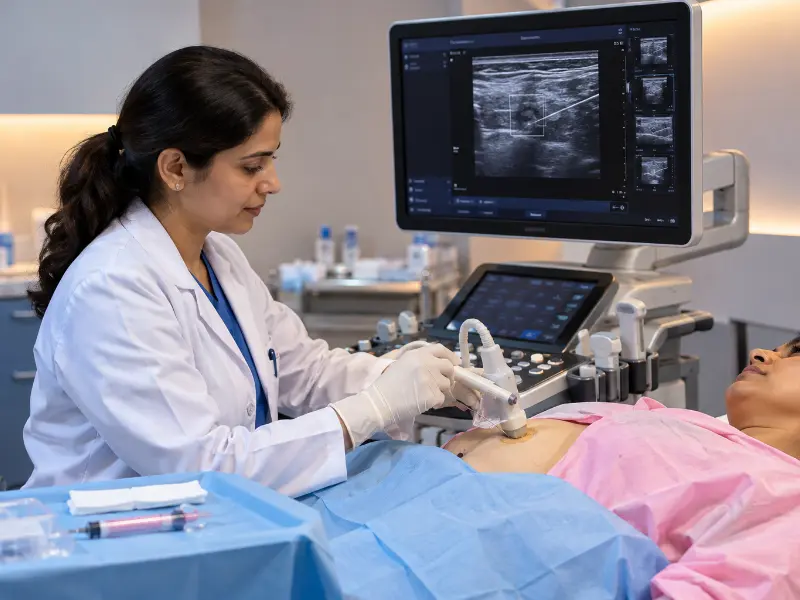
A Guided Biopsy is a minimally invasive diagnostic procedure that uses real-time ultrasound imaging to guide a core needle into a selected lesion. As a result, the radiologist can obtain tissue samples from the targeted area for histopathological examination.
In particular, ultrasound-guided tissue sampling may be used for appropriate breast lumps, lymph nodes, liver lesions, thyroid lesions and selected soft tissue masses. Moreover, the procedure allows the radiologist to visualize the target during needle placement.
At Henotic Diagnostics, Kharghar, appropriate core needle tissue sampling procedures are performed using high-resolution ultrasound imaging. Depending on the clinical requirement, local anesthesia may be used to improve patient comfort.
Therefore, the collected tissue can be submitted for pathological evaluation when a treating physician requires additional information about a lesion. The exact procedure depends on the location, size and characteristics of the target.
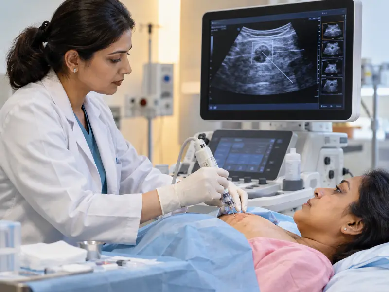
A Guided Biopsy combines live ultrasound imaging with core needle tissue sampling. During the procedure, the radiologist uses real-time sonography to identify the target and monitor the needle as it approaches the selected area.
Unlike sampling performed without imaging guidance, ultrasound provides continuous visualization during an appropriate procedure. Therefore, the radiologist can assess the target and needle position throughout the sampling process.
Furthermore, the collected tissue may preserve important tissue architecture. Consequently, the pathology laboratory can examine the sample using histopathological methods when clinically indicated.
For example, tissue sampling may be considered when a breast lesion, lymph node, liver lesion, thyroid lesion or soft tissue mass requires further diagnostic evaluation.
For patient information about breast tissue sampling, visit Mayo Clinic Breast Biopsy Overview .
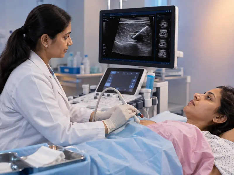
A Guided Biopsy provides tissue for laboratory evaluation when imaging or clinical findings identify a suspicious or indeterminate lesion. In addition, the collected sample may help the treating team understand the nature of the tissue.
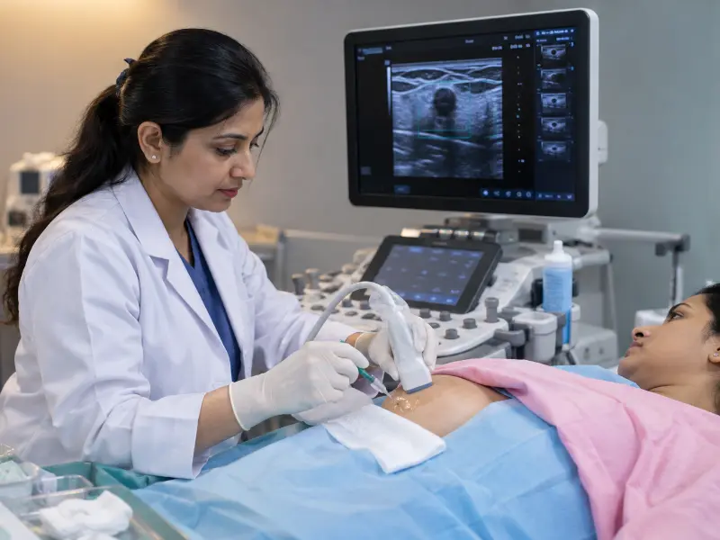
A treating physician may recommend a Guided Biopsy when physical examination, ultrasound, mammography, MRI or another diagnostic assessment identifies a lesion that requires tissue diagnosis.
For instance, selected breast lesions, enlarged lymph nodes, suspicious liver or thyroid lesions and appropriate soft tissue masses may require further evaluation. However, the decision to perform targeted tissue sampling depends on the clinical history, imaging findings and medical assessment.
In addition, the treating physician may request tissue evaluation when imaging alone cannot provide enough information for the clinical question. Therefore, patients should follow the recommendation of their doctor and radiologist.
Image-guided tissue sampling may be recommended when a physician requires tissue for histopathological evaluation of a suspicious or indeterminate lesion. In such cases, the selected imaging method depends on the location and characteristics of the target.
For appropriate accessible lesions, ultrasound-guided core sampling provides real-time visualization during needle placement. Additionally, ultrasound can help the radiologist assess the target and nearby structures during the procedure.
The final decision regarding the procedure should be made by the treating medical team based on the patient’s clinical and imaging findings.
For professional information regarding ultrasound-guided procedures, visit AIUM Practice Parameters for Ultrasound-Guided Procedures .
Ultrasound provides real-time visualization of many superficial and accessible lesions. Therefore, it can help the radiologist monitor the target and needle position during an appropriate procedure.
Moreover, ultrasound-guided tissue sampling allows continuous imaging while the needle is directed toward the selected area. This approach can help the radiologist account for nearby anatomical structures.
Another advantage is that diagnostic ultrasound does not use ionizing radiation. However, the most appropriate guidance technique depends on the location and characteristics of the lesion.
As a result, ultrasound may be selected when real-time sonographic visualization is clinically suitable.
A Guided Biopsy is generally scheduled after referral or recommendation from the treating physician or specialist. Before the appointment, patients should provide relevant medical information.
Additionally, follow any specific preparation instructions provided by your treating doctor or radiologist.
Your doctor may recommend tissue sampling when imaging or clinical examination identifies a breast lump, suspicious mammographic finding, enlarged lymph node, focal liver lesion, thyroid lesion or soft tissue mass.
For example, a lesion that requires microscopic evaluation may be referred for core tissue sampling when clinically appropriate. However, not every lump or abnormal imaging finding requires a biopsy.
In addition, another cell-based diagnostic procedure may sometimes be appropriate. Explore our Guided FNAC Services for related information.
Henotic Diagnostics provides selected ultrasound-guided core sampling procedures according to the target anatomical site and clinical requirement.
Meanwhile, the specific sampling method depends on the lesion, its location and the clinical question being investigated.
A Guided Biopsy provides tissue that may help a pathology laboratory evaluate a range of conditions. Importantly, the final diagnosis depends on tissue findings together with the patient’s clinical and imaging information.
Consequently, tissue results are interpreted together with the patient’s history and imaging findings rather than being considered in isolation.
Image-guided tissue sampling combines imaging visualization with needle-based tissue collection. When ultrasound is clinically suitable, this approach provides real-time visualization during the sampling process.
Therefore, an ultrasound approach may be useful for selected lesions that can be adequately visualized with sonography.
Preparation for a Guided Biopsy depends on the target location, medical history and procedure planned by the radiologist. Patients should provide information about medications, allergies and relevant medical conditions.
During an ultrasound-guided procedure, the patient is positioned so that the target lesion can be visualized. Next, the skin is cleaned using appropriate sterile precautions, and local anesthesia may be administered when clinically appropriate.
Under continuous ultrasound visualization, a core needle is directed toward the target and tissue samples are collected. Subsequently, the samples are placed in appropriate containers and sent for pathological evaluation when required.
After sampling, the entry site is cleaned, compressed and covered with an appropriate dressing. Finally, patients receive post-procedure instructions according to the procedure performed and their individual clinical circumstances.
Henotic Diagnostics provides transparent pricing for selected Guided Biopsy and core tissue sampling procedures. However, the final cost may depend on the anatomical site, procedure complexity, pathology requirements and additional laboratory testing.
| Procedure | Approximate Cost (₹) |
|---|---|
| USG-Guided Breast Core Tissue Sampling | ₹3,500 – ₹8,500 |
| Lymph Node / Soft Tissue Core Sampling | ₹3,000 – ₹8,000 |
| Visceral / Deep Mass Tissue Sampling | ₹4,500 – ₹8,000 |
For this reason, please confirm the applicable procedure cost before booking. Pricing can vary according to the required tissue sampling procedure and laboratory investigations.
Core tissue sampling and FNAC are different sampling techniques. Core sampling obtains small cylinders of tissue that preserve tissue architecture, whereas FNAC uses a fine needle to collect individual cells for cytological examination.
| Feature | Core Tissue Sampling | FNAC |
|---|---|---|
| Sample | Tissue core | Individual cells |
| Laboratory Evaluation | Histopathology | Cytology |
| Tissue Architecture | Preserved in the tissue core | Not obtained as an intact tissue core |
| Additional Testing | May be possible when clinically appropriate | Depends on the cells obtained and clinical requirement |
Nevertheless, the appropriate technique depends on the lesion, clinical question, imaging findings and recommendation of the treating medical team. For additional information about ultrasound-guided procedures, visit AIUM Practice Parameters for Ultrasound-Guided Procedures .
A Guided Biopsy is generally performed under local anesthesia to numb the targeted area. As a result, patients may feel pressure or mild discomfort during tissue sampling.
Real-time ultrasound imaging allows the radiologist to visualize the target lesion and direct the core needle toward the selected tissue. Therefore, the needle position can be monitored during the sampling process.
The tissue sampling and ultrasound guidance may take approximately 20 to 30 minutes. However, the overall appointment may take about 45 minutes, including preparation and post-procedure observation.
FNAC collects individual cells for cytological examination, whereas core tissue sampling obtains small tissue cores that preserve tissue architecture for histopathological evaluation.
The timing of the final report depends on the laboratory analysis and the tests requested. In addition, histopathology and further investigations may take several business days.
Bring your doctor’s prescription or referral, previous ultrasound, mammography, CT or MRI reports and images when available. Also, inform the radiologist about regular medications, especially blood-thinning medicines.
|
|
||||||
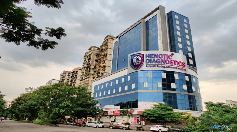
Henotic Diagnostics in Kharghar provides diagnostic imaging and selected interventional procedures, including ultrasound-guided tissue sampling when clinically appropriate. Additionally, the centre focuses on patient privacy, procedure preparation and diagnostic coordination.
The facility supports appropriate imaging-based procedures using ultrasound visualization. Consequently, patients can discuss their procedure requirements with the diagnostic team before scheduling.
Address: Second floor, Millennium Empire, Business Park, Plot No 47, D Mart Rd, Sector 15, Kharghar, Navi Mumbai, Panvel, Maharashtra 410210.
Phone: 088793 27184
⭐⭐⭐⭐⭐
“Extremely smooth and reassuring experience for my breast Guided Biopsy! Dr. Pratibha Patil explained the local numbing process and guided core sampling with complete care.”
⭐⭐⭐⭐⭐
“Very fast Guided Biopsy appointment. The ultrasound procedure report was issued promptly and the biopsy core results came back quickly.”
Henotic Diagnostics provides Guided Biopsy and selected ultrasound-guided tissue sampling services in Kharghar. Our diagnostic team focuses on appropriate imaging guidance, patient comfort, privacy and coordination of tissue diagnosis with the treating medical team.
Furthermore, patients can discuss preparation requirements and appointment details with the diagnostic team before the procedure.
If your doctor has recommended a Guided Biopsy or ultrasound-guided tissue sampling, contact Henotic Diagnostics to discuss the procedure and appointment requirements.
Call 088793 27184 to book your appointment slot.
Alternatively, explore our full range of diagnostic ultrasound services online.
|
See what our patients have to say about their diagnostic experience with us.
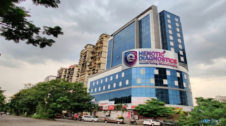
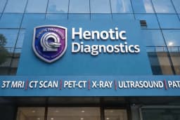

"Excellent service! The staff is very cooperative and the facility is spotless. Got my MRI done smoothly."
 Posted on Google
"State-of-the-art equipment and highly professional doctors. Reports were delivered on time via WhatsApp. Highly recommended."
Posted on Google"Very convenient location in Kharghar. The home collection team was punctual and well-trained. Great experience overall."
Posted on Google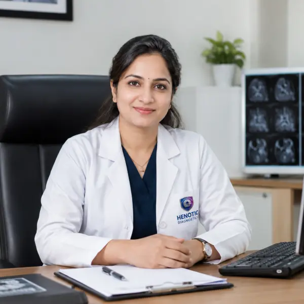

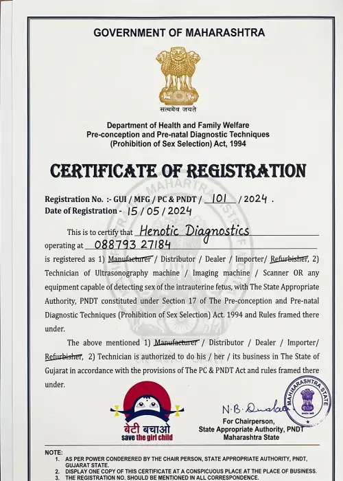

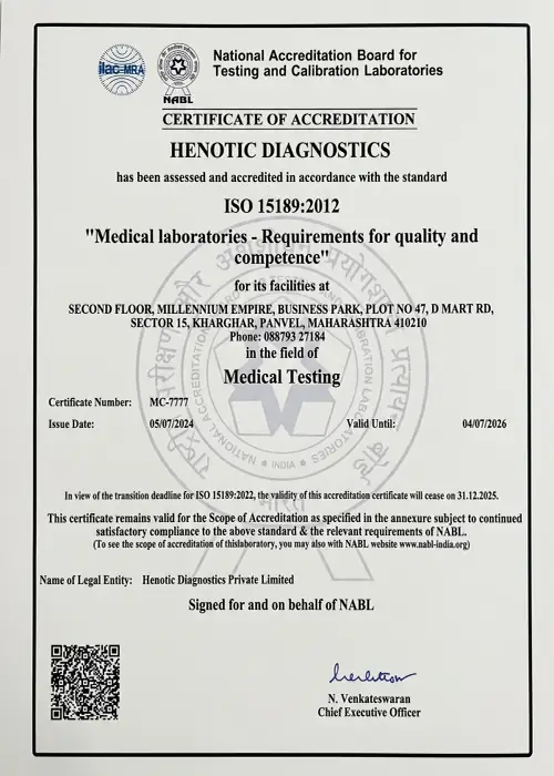

 View on Google Maps