
When your physician or surgeon orders a Magnetic Resonance Imaging (MRI) scan, you may encounter options between a 1.5 Tesla (1.5T) and a 3.0 Tesla (3.0T) scanner. Understanding what these numbers signify can help you choose the ideal diagnostic scan for your specific condition.
What Does 'Tesla' Mean in MRI Technology?
In physics and diagnostic radiology, Tesla (T) is the International System (SI) unit measuring magnetic field strength. One Tesla equals approximately 10,000 Gauss. For perspective, the Earth's natural magnetic field is roughly 0.5 Gauss. Thus, a 1.5T scanner creates a magnetic field approximately 30,000 times stronger than Earth, while a 3.0T scanner generates a field 60,000 times stronger.
The primary benefit of a stronger magnetic field is a higher Signal-to-Noise Ratio (SNR). In practical terms, 3.0T offers double the diagnostic signal strength of 1.5T, allowing radiologists to capture micro-anatomical structures with ultra-fine millimeter slice thicknesses.
Key Differences: 1.5T vs 3.0T MRI Comparison
| Feature | 1.5 Tesla MRI | 3.0 Tesla MRI |
|---|---|---|
| Magnetic Field Strength | 15,000 Gauss | 30,000 Gauss (2x) |
| Spatial Resolution | Standard High Resolution (~1.5mm) | Ultra-High Resolution (sub-millimeter <0.5mm) |
| Average Scan Duration | 25 – 45 Minutes | 15 – 25 Minutes (up to 40% faster) |
| Best Clinical Applications | General screening, spine, abdomen, patients with older implants | Brain stroke, epilepsy, micro-tumors, pituitary, MSK cartilage, prostate multiparametric MRI |
| Radiation Exposure | Zero Ionizing Radiation | Zero Ionizing Radiation |
When is a 3.0T MRI Essential?
While 1.5T scanners provide dependable diagnostic clarity for routine screening, 3.0T MRI has become the global gold standard for complex diagnostic challenges:
- Neuroimaging & Stroke Detection: 3.0T diffusion-weighted imaging (DWI) detects acute ischemic stroke within minutes of symptom onset, identifying micro-infarcts and cortical dysplasia that 1.5T might miss.
- Musculoskeletal & Sports Medicine: Exceptional visualization of sub-millimeter labral tears in the hip or shoulder, cruciate ligament bundle tears in the knee, and early articular cartilage erosion.
- Multiparametric Prostate MRI (mpMRI): High-resolution T2-weighted and dynamic contrast-enhanced imaging using PIRADS scoring without requiring an invasive endorectal coil.
- Epilepsy & Brain Tumor Margins: Pinpointing epileptogenic cortical malformations, hippocampal sclerosis, and microscopic neoplastic margins for neurosurgical precision.
Patient Comfort, Scan Time & Claustrophobia
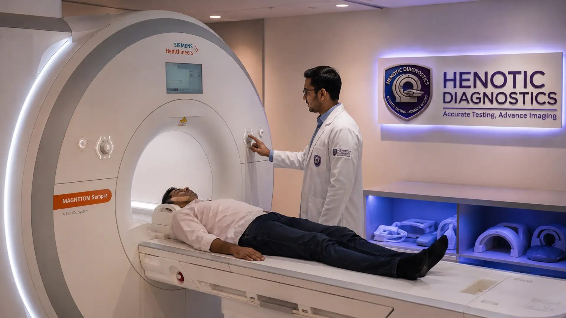
Modern 3.0T systems, such as Henotic Diagnostics' wide-bore 70cm configuration, significantly diminish claustrophobia. Older traditional scanners featured narrow 60cm tunnels, creating anxiety for patients. A 70cm wide-bore system allows ample headroom, calm ambient lighting, and high-fidelity noise-reduction headsets.
Furthermore, reduced scan durations (completing a comprehensive cervical spine or brain protocol in under 15 minutes) reduce motion artifacts and make the experience much easier for elderly patients or children.
Implants and Safety Considerations
Safety is the foremost priority in any MRI suite. Before entering the MRI room (Zone IV), patients must undergo an exhaustive screening checklist:
- Cardiac Pacemakers & Defibrillators: Modern MRI-conditional pacemakers require programming into 'MRI mode' prior to scanning.
- Orthopedic Hardware: Most modern titanium pins, plates, and joint replacements are MRI-safe; however, our technologists verify manufacturer documentation.
- Metallic Foreign Bodies: Patients with a history of metal grinding or shrapnel exposure undergo a quick pre-scan screening orbit X-ray to rule out ocular metallic fragments.
Clinical Takeaway
If your referring neurologist, neurosurgeon, or orthopedic specialist requests precise visualization of small nerve roots, cartilage, or brain parenchyma, opting for a 3.0T wide-bore MRI scan ensures definitive diagnostic accuracy the first time, preventing inconclusive results and repeat imaging.
Frequently Asked Questions
Is a 3.0T MRI always better than a 1.5T MRI?
3.0T provides twice the signal-to-noise ratio, resulting in sharper detail and faster scan times for brain, spine, microvascular, and complex joint cartilage evaluations. However, 1.5T remains clinically excellent for general body imaging and is often preferred for patients with certain older conditional metallic implants or pacemakers.
Does a 3.0T MRI scan expose me to radiation?
No. Magnetic Resonance Imaging (MRI) uses powerful magnetic fields and radiofrequency waves. It uses zero ionizing radiation, making it completely safe from radiation risks at both 1.5T and 3.0T strengths.
How long does a 3T MRI scan take compared to a 1.5T scan?
Because 3.0T systems acquire diagnostic signals much faster, scan durations are frequently reduced by 30% to 50%, often completing a detailed sequence in 12–20 minutes instead of 30–45 minutes.
Need This Diagnostic Scan?
Henotic Diagnostics provides same-day appointments, 3.0T wide-bore imaging, and NABL-certified reporting across Mumbai & Navi Mumbai.