
Cardiovascular diseases remain the leading cause of mortality in India. Because heart conditions present through different physiological mechanisms — electrical arrhythmias, structural valve defects, or exercise-induced ischemia — distinct diagnostic tools are required to build a complete clinical picture.
The Non-Invasive Cardiac Diagnostic Triad
Cardiologists rely on three foundational non-invasive investigations to evaluate heart health:
1. Resting 12-Lead ECG: Electrical Rhythms & Immediate Alerts
The Electrocardiogram (ECG) records the electrical voltage generated by your heart muscle during each cardiac cycle. Electrodes placed across your chest, wrists, and ankles capture electrical impulses in milliseconds.
- Best for: Detecting acute myocardial infarction (heart attack via ST-elevation), cardiac arrhythmias (atrial fibrillation, tachycardia, bradycardia), and heart block.
- Limitation: A normal resting ECG cannot rule out underlying coronary artery disease or future heart attack risks.
2. 2D Echocardiography: Heart Architecture & Ejection Fraction
A 2D Echo is a non-invasive ultrasound examination of the moving heart. By placing a specialized acoustic transducer on the chest wall, the cardiologist visualizes live real-time videos of cardiac chambers and moving valves.
- Best for: Measuring Left Ventricular Ejection Fraction (LVEF), detecting heart enlargement (cardiomegaly), identifying valve stenosis or regurgitation, assessing pericardial effusion, and identifying regional wall motion abnormalities (RWMA) from past silent attacks.
- Zero Radiation: Uses pure ultrasound waves, making it repeatable and completely safe.
3. TMT Stress Test: Uncovering Exertional Ischemia
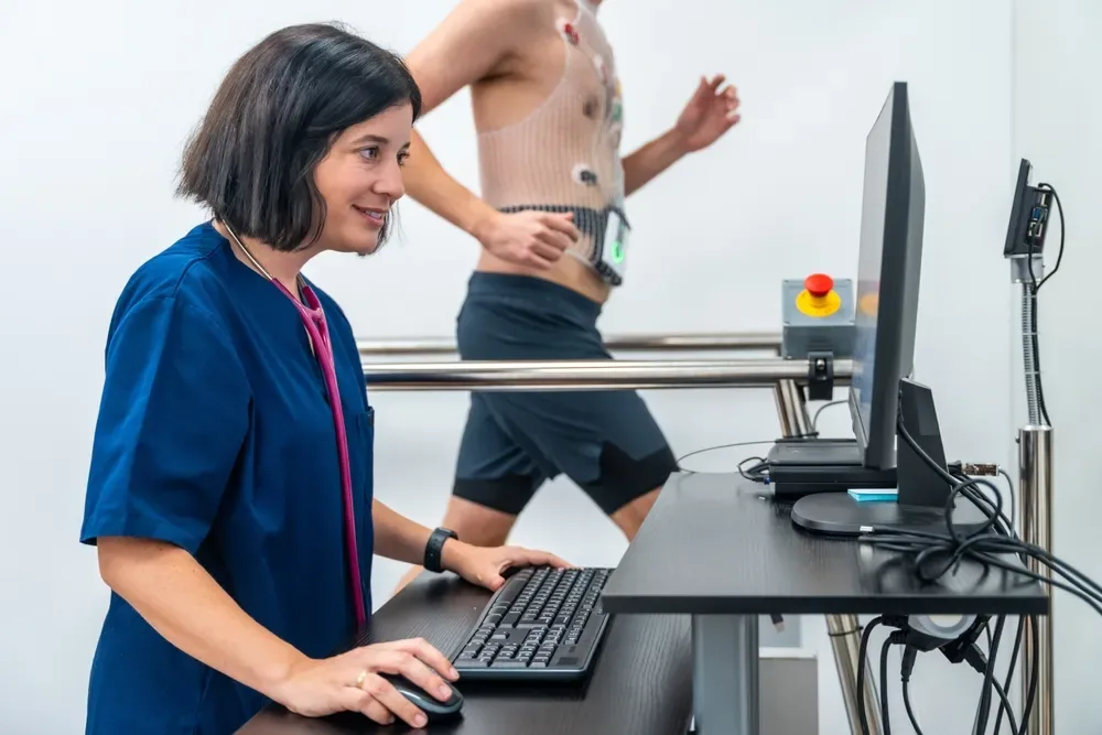
The Treadmill Test (TMT) monitors your ECG, heart rate, and blood pressure continuously while you walk and run on a calibrated motorized treadmill following the standardized Bruce Protocol.
- Best for: Diagnosing inducible coronary artery ischemia. When your workload increases, the heart muscle requires more oxygenated blood. If narrowed coronary arteries cannot deliver enough blood, classic ST-segment depressions appear on the stress monitor before you even feel chest tightness.
Which Test Should You Book?
- If you experience acute, sudden crushing chest pain: Seek emergency care immediately for an instant emergency ECG and Troponin blood test.
- If you have hypertension, diabetes, or shortness of breath on climbing stairs: Book a 2D Echocardiography + Resting ECG.
- If you are aged 40+, starting a vigorous gym fitness routine, or experiencing exertion-induced chest heaviness: Book a TMT Stress Test.
Frequently Asked Questions
Can an ECG be completely normal even if I have heart blockages?
Yes. A resting 12-lead ECG records electrical signals while you are lying calmly on an exam bed. In stable coronary artery disease, blood flow at rest is often adequate to maintain normal electrical patterns. Significant blockages (70%+) often only manifest when the heart is challenged during a TMT stress test.
What does Ejection Fraction (EF) on a 2D Echo mean?
Ejection Fraction measures the percentage of blood pumped out of your left ventricle with each contraction. A normal healthy EF ranges between 55% and 70%. Values below 50% indicate reduced myocardial pumping capability or heart failure.
How should I dress for a TMT (Treadmill Test)?
Wear comfortable, loose workout attire and supportive athletic running shoes. Avoid heavy meals for 2 to 3 hours before the test, and consult your cardiologist regarding whether to withhold beta-blocker medications on test morning.
Need This Diagnostic Scan?
Henotic Diagnostics provides same-day appointments, 3.0T wide-bore imaging, and NABL-certified reporting across Mumbai & Navi Mumbai.